
Page 1538 - Hematology_ Basic Principles and Practice ( PDFDrive )
P. 1538
Chapter 85 T-Cell Lymphomas 1365
variable numbers of admixed inflammatory cells often expanding into similar to those of MF. However, the cellular infiltrates in SS are more
adnexal structures (hair follicles and sweat glands). Epidermal exocy- often monotonous, and epidermotropism may be absent.
tosis of single or small clusters of neoplastic cells is a characteristic Lymph node involvement initially involves the paracortical
finding (Fig. 85.18A, C, and D). The presence of Pautrier microab- regions. Progression is associated with small-to-large clusters of atypi-
scesses, defined as four or more atypical lymphocytes arranged in an cal cells with preserved nodal architecture, followed by partial or
aggregate in the epidermis, is classic but is seen in only a minority of total effacement of the node by neoplastic cells. Visceral involve-
cases. Reticular fibroplasia of the papillary dermis is also a common ment is a late clinical feature. Variable peripheral blood involvement
finding. Tumor-stage lesions demonstrate a more diffuse, superficial, can be demonstrated in all stages of skin disease, although it is most
and deep dermal infiltrate with fewer reactive cells and an absence of prevalent in patients with tumor or erythrodermic presentations.
epidermotropism (Fig. 85.18B, E, and F). The malignant T-cell clone Patients with SS present with large numbers of circulating neoplastic
often evolves into large-cell morphology during tumor progression, cells, typically more than 1000 cells/mL (B2), but a lower count
although rare cases show large-cell morphology from the early patch may be noted initially (B1). Bone marrow biopsy results are typically
lesions. The presence of large cells in a skin lesion should be distin- negative. However, some involvement may be detected by flow
guished from “large-cell transformation,” which displays rapid skin, cytometry in many cases of SS or advanced MF but rarely influences
node, and visceral progression, is refractory to treatment, and is management outside an investigational setting. A staging bone
associated with poor survival. The histologic features in SS may be marrow aspirate and biopsy is not recommended, unless unexplained
cytopenias are seen.
+
+
+
+
The malignant cells are typically CD3 , CD4 , CD45RO , CD8 ,
+
and CD30 by immunohistochemical analysis. CD7 is not expressed
by cells from the early disease stages. More aggressive variants and
advanced forms of CTCL may have cells with multiple pan–T-cell
antigen deletions, especially CD2, CD5, and even CD4. TCR genes
are clonally rearranged and can be documented in most cases by
Southern blotting or polymerase chain reaction assays when a suffi-
cient malignant infiltrate exists.
Analysis of peripheral blood may reveal an elevated LDH level,
mostly in patients with high-blood–burden SS and bulky advanced
MF. Eosinophilia and hypergammaglobulinemia are occasionally
observed in advanced SS. In addition, several reports of SS and other
CTCL variants have been reported to be associated with B-cell
lymphoproliferative conditions, or small peripheral B-cell clones or
monoclonal gammopathy (PMID: 19481294). Elevated serum β 2 -
microglobulin and IL-2 receptor levels have also been observed in
advanced cases.
Imaging studies for classic MF/SS are generally of modest utility.
Computed tomographic scans of the chest, abdomen, or pelvis should
be reserved for patients with SS, nodal involvement, or CTCL vari-
ants. In an investigational setting, electron microscopy, cytogenetics,
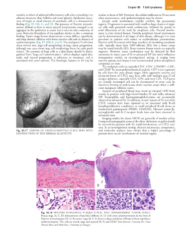
Fig. 85.17 LESIONS OF GRANULOMATOUS SLACK SKIN WITH and molecular analyses have shown that a higher percentage of
DESTRUCTION OF THE DERMAL ELASTICITY. patients have occult involvement of internal organs.
A B
C C D E F
Fig. 85.18 MYCOSIS FUNGOIDES, PLAQUE STAGE, AND TRANSFORMED TUMOR STAGE.
Plaque stage (A, C, D) demonstrates a band-like infiltrate (A, C) with some epidermotropism in the form of
Pautrier microabscesses (D). In the tumor stage (B, E, F) there is a deep and dense infiltrate without significant
+
epidermotropism. The cells are mostly large and atypical (E, F) and CD30 (not shown). (Courtesy Drs. Vesna
Petrovic-Rosic and Mark Racz, University of Chicago.)