
Page 1537 - Hematology_ Basic Principles and Practice ( PDFDrive )
P. 1537
1364 Part VII Hematologic Malignancies
can be associated with clonal T-lymphocytic infiltration. In general, origin. Although the typical course is usually indolent, spanning
patients older than 40 years with a more generalized cutaneous decades, approximately 15% of patients develop MF, HL, or NHL
involvement and a chronic course are more likely to develop associ- during their lifetime. A direct link between LyP, CTCL, and HL was
ated MF. No cases of MF have been reported in children with alopecia demonstrated in a patient with the three lymphoproliferative disor-
mucinosa, although a few reports of HL have been reported in ders arising from a common T-cell clone, as shown by TCR gene
children with follicular mucinosis. Patients with folliculotropic MF studies.
and follicular mucinosis are reported to have a worse prognosis, stage
for stage, which may be caused by inability of topical treatment to
penetrate to the deeper layers of the process. Pagetoid Reticulosis
Pagetoid reticulosis (i.e., Woringer-Kolopp disease) is a rare condition
Lymphomatoid Papulosis affecting young adults. It typically manifests with a solitary, hyper-
keratotic, often verrucous plaque on the lower limb. Biopsy results
Lymphomatoid papulosis (LyP) is characterized by recurrent crops of show atypical cerebriform lymphocytes with a perinuclear halo almost
self-healing, red-brown, centrally necrotic, asymptomatic papules and exclusively localized within the intraepidermal compartment. Extra-
+
nodules (Fig. 85.15). This entity represents 10%–15% of all CTCL cutaneous dissemination is exceedingly rare. Most cases have a CD8
+
+
cases. Patients may have a few lesions or more than 100 at a time. phenotype, although CD4 cases or double-negative (CD4 /CD8)
+
Histologic evaluation reveals an atypical CD4 lymphocytic infiltrate cases have been reported. Frequently the tumor cells express CD30,
with a variable mixed inflammatory infiltrate (Fig. 85.16). These may but TCR gene rearrangement study results are often negative. Whether
be primarily small cerebriform cells similar to those seen in MF (type pagetoid reticulosis should be considered a localized form of MF or
+
B), but most often there are larger CD30 cells with prominent a reactive pseudomalignant process is debatable. Although most cases
nucleoli resembling Reed-Sternberg cells (type A). A third variety of have an indolent protracted course, generalized and sometimes
+
LyP (type C) with sheets of anaplastic large cells resembling CD30 aggressive variants have been reported. Cases presenting with a soli-
+
large-cell lymphoma (CD30 LCL) has also been reported. Lately tary lesion are extremely indolent and could be considered a reactive
other subtypes have been reported. Type D, commonly seen in the or pseudolymphomatous process. At the opposite end of the
pediatric population, is characterized by pagetoid or highly epider- spectrum there are patients with extensive ulcerative plaques formerly
motropic small atypical lymphocytes with CD8 expression. Such known as generalized pagetoid reticulosis or Ketron-Goodman disease
+
cases may be difficult to distinguish from pityriasis lichenoides acuta that are now diagnosed as the CD8 aggressive intraepidermal T-cell
(PMID: 22688398). lymphoma.
Type E LyP is characterized by hemorrhagic and necrotic lesions
often showing evidence of vasculitis on histological evaluation
(PMID: 23026936). Granulomatous Slack Skin
The prognosis of these new subtypes is not different to the other
subgroups. TCR gene rearrangement studies demonstrate a clonal In granulomatous slack skin syndrome, an extremely rare disorder,
+
clonal CD4 T cells elicit a reactive granulomatous response that
destroys the elastic fibers, rendering skin slack, fibrotic, and inelastic
(Fig. 85.17). Changes characteristic of MF are often found within
the epidermis and papillary dermis, and the reticular dermis contains
numerous histiocytes with multinucleated giant cells and elasto-
phagocytosis. Some patients with granulomatous MF do not have
destruction of the elastic fibers with slack skin changes. The differ-
ential diagnosis includes sarcoidosis and tuberculoid leprosy. An
increased incidence of HL has been reported in this patient
population.
Laboratory Manifestations
The gold standard in the diagnosis of MF/SS is light microscopic
examination of a skin biopsy specimen. Characteristic findings
include a band-like infiltrate involving the papillary dermis contain-
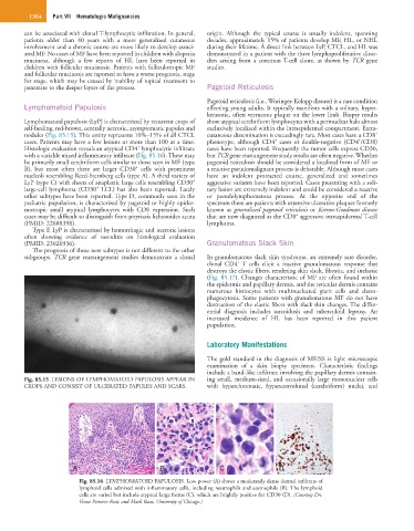
Fig. 85.15 LESIONS OF LYMPHOMATOID PAPULOSIS APPEAR IN ing small, medium-sized, and occasionally large mononuclear cells
CROPS AND CONSIST OF ULCERATED PAPULES AND SCARS. with hyperchromatic, hyperconvoluted (cerebriform) nuclei, and
A B C D
Fig. 85.16 LYMPHOMATOID PAPULOSIS. Low power (A) shows a moderately dense dermal infiltrate of
lymphoid cells admixed with inflammatory cells, including neutrophils and eosinophils (B). The lymphoid
cells are varied but include atypical large forms (C), which are brightly positive for CD30 (D). (Courtesy Drs.
Vesna Petrovic-Rosic and Mark Racz, University of Chicago.)