
Page 172 - Hematology_ Basic Principles and Practice ( PDFDrive )
P. 172
130 Part II Cellular Basis of Hematology
TABLE Other Adhesion Receptors
12.3
Name Other Name Expressed by Ligand Function(s)
Cadherins EC, many other cells Homotypic binding Formation of EC junctions
GPlb/IX/V Platelets vWF Platelet adhesion to ECM under shear
CD36 GPIV Platelets, many other cells Collagens, TSP Platelet adhesion to ECM
CD44 Leukocytes, other cells Hyaluronan, serglycin Lymphopoiesis, lymphocyte activation
DC-SIGN Dendritic cells Mannosylated glycans, other glycans Regulate T-cell–dendritic cell interactions,
recognize pathogens
NK cell receptors NK cells MHC molecules Recognition of virus-infected or other
foreign cells
DC-SIGN, Dendritic cell-specific ICAM-3 grabbing nonintegrin; MHC, major histocompatibility complex; NK, natural killer. For other abbreviations, see Table 12.1
footnotes.
TABLE Selectins
12.4
Name Other Name Expressed by Ligand Ligands Expressed by Function(s)
P-selectin CD62P GMP-140 Thrombin-activated PSGL-1, GPlbα Leukocytes, platelets Leukocyte adhesion
PADGEM platelets and ECs, to activated ECs
cytokine-activated ECs and platelets
E-selectin CD62E ELAM-1 Cytokine-activated ECs PSGL-1, other sialylated Leukocytes Leukocyte adhesion to
and fucosylated GPs activated ECs
L-selectin CD62L LECAM-1 LAM-1 Leukocytes PSGL-1, also GlyCAM-1, Leukocytes, ECs or Leukocyte adhesion to
CD34, and other lymph nodes other leukocytes;
mucins on ECs of lymphocyte homing
lymph nodes to lymph nodes
The selectins bind to sialylated, fucosylated, and (in some cases) sulfated oligosaccharides on specific glycoproteins, of which only some have been identified.
EC, Endothelial cell; ELAM-1, endothelial leukocyte adhesion molecule-1; Gly-CAM-1, glycosylation-dependent cell adhesion molecule-1; GMP-140, granule membrane
protein-140; LAM-1, leukocyte adhesion molecule-1; LECAM-1, leukocyte endothelial cell adhesion molecule-1; PADGEM, platelet activation-dependent granule external
membrane protein; PSGL-1, P-selectin glycoprotein ligand-1. For other abbreviations, see Table 12.1 footnotes.
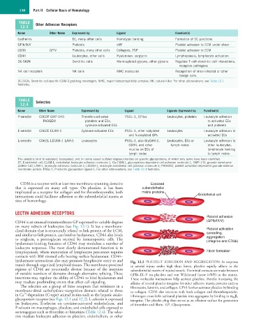
CD36 is a receptor with at least two membrane-spanning domains Exposed
that is expressed on many cell types. On platelets, it has been subendothelial
implicated as a receptor for collagen and for thrombospondin; both matrix proteins
interactions could facilitate adhesion to the subendothelial matrix at Endothelial cell
sites of hemorrhage.
LECTIN ADHESION RECEPTORS
Platelet adhesion
CD44 is an unusual transmembrane GP expressed to variable degrees (GPIb/IX/V)
on many subsets of leukocytes (see Fig. 12.1). It has a membrane-
distal domain that is structurally related to link protein of the ECM, Platelet activation
and similar to link protein, can bind to hyaluronan. CD44 also binds spreading,
aggregation
to serglycin, a proteoglycan secreted by hematopoietic cells. The (integrins and CD36)
hyaluronan-binding function of CD44 may modulate a number of
leukocyte responses. The most clearly demonstrated function is in
lymphopoiesis, where maturation of lymphocyte precursors requires Fibrin formation
contacts with BM stromal cells bearing surface hyaluronan. CD44–
hyaluronate interactions also may promote lymphocyte entry to and Fig. 12.2 PLATELET ADHESION AND AGGREGATION. In response
transit through organized lymphoid tissues. The membrane-proximal to arterial injury under high shear forces, platelets rapidly adhere to the
regions of CD44 are structurally diverse because of the insertion subendothelial matrix of injured vessels. The initial contacts are made between
of variable numbers of domains through alternative splicing. These GPIb–IX–V on platelets and von Willebrand factor (vWF) in the matrix.
insertions may regulate the ability of CD44 to bind hyaluronan and These molecular interactions help activate platelets, thereby increasing the
may mediate postbinding events that affect cell signaling. affinity of several platelet integrins for other adhesive matrix proteins such as
The selectins are a group of three receptors that terminate in a fibronectin, laminin, and collagen. GPVI further activates platelets by binding
membrane-distal carbohydrate-recognition domain related to those to collagen. CD36 also interacts with both collagen and thrombospondin.
2+
in Ca -dependent (C-type) animal lectins such as the hepatic asialo- Fibrinogen cross-links activated platelets into aggregates by binding to α IIb β 3
glycoprotein receptor (see Figs. 12.1 and 12.2). L-selectin is expressed integrins. The platelet plug then serves as an efficient surface for generation
on leukocytes, E-selectin on cytokine-activated endothelium, and of thrombin and fibrin. GP, Glycoprotein.
P-selectin on macrophages, platelets, and endothelial cells exposed to
secretagogues such as thrombin or histamine (Table 12.4). The selec-
tins mediate leukocyte adhesion to platelets, endothelium, or other