
Page 175 - Hematology_ Basic Principles and Practice ( PDFDrive )
P. 175
Chapter 12 Cell Adhesion 133
unstimulated platelets to home to the site of vascular injury and then
be activated by locally generated mediators.
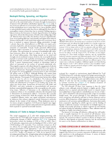
CD28 B7-1/B7-2
Neutrophil Rolling, Spreading, and Migration
Near sites of extravascular bacterial infections, neutrophils first tether to LFA-1 ICAM 1,2,3
and roll on the endothelial surface of venules through the interactions
of selectins with cell-surface carbohydrate ligands (Fig. 12.3). Little, if CD-2 Antigen-
any, leukocyte adhesion occurs in nearby arterioles. Neutrophil rolling T cell LFA-3 presenting
on the endothelium occurs under shear forces, just as platelets adhere cell
to subendothelial matrix under shear forces, although the shear flow in TCR/CD3
postcapillary venules is lower than that in arterioles. Rolling requires a
balance between the formation of selectin–ligand bonds at the leading MHC
edge of the cell and the dissociation of bonds at the trailing edge of the CD4/CD8
cell. Whereas shear forces affect the lifetimes of selectin–ligand bonds,
lower forces prolong lifetimes (catch bonds) and higher forces shorten
lifetimes (slip bonds). Catch bonds help explain why a minimum shear Fig. 12.4 ADHESION BETWEEN T LYMPHOCYTES AND ANTIGEN-
force is required to support leukocyte rolling, particularly through PRESENTING CELLS. The initial contact is mediated by the TCR, or CD3,
L-selectin. Just as the initial adhesion to vWF does not require prior which binds with low affinity but high specificity to a specific antigen pre-
activation of platelets, selectin-mediated rolling does not require prior sented by a MHC molecule. Additional contacts, also of low affinity, are
activation of neutrophils. Instead, locally generated inflammatory between CD4 (on helper cells) or CD8 (on cytotoxic cells) and MHC, and
mediators induce expression of E- or P-selectin on the endothelial between costimulatory molecules CD2 and LFA-3 and CD28 and CD80
cell surface. The requirement for activation of endothelial cells rather (B7-1) and CD86 (B7-2). These interactions signal the T cell to transiently
than leukocytes allows the latter to adhere to vessels only at the increase the affinity of the β 2 integrin LFA-1 for ICAM-1 on the antigen-
site of vessel inflammation. After being situated on the vessel wall presenting cell. These bonds strengthen adhesion, and the costimulatory
through selectin-mediated contacts, however, the neutrophils become molecules transduce sustained and additional signals to the T cell that cause
exposed to activators such as platelet-activating factor, a phospholipid increased gene transcription, proliferation, and cytokine secretion. Not shown
signaling molecule, activated complement proteins, and interleukin-8 is the redistribution of these adhesion molecules into different regions of the
(IL-8), a potent chemoattractant cytokine or chemokine, both of contact zone as adhesion strengthens. Additional signals result from binding
which are presented on the surface of activated endothelial cells. of β 1 integrins on the T cell to adhesive proteins in the extracellular matrix.
These signals cooperate with others directed by engagement of selectin LFA-3, Leukocyte function-associated antigen-3; MHC, major histocompat-
ligands and promote very slow rolling of neutrophils on the surface of ibility complex; TCR, T-cell receptor.
activated endothelium. Neutrophil activation increases the affinity of
β 2 integrins for immunoglobulin counterreceptors on the endothelial
cell surface such as ICAM-1. Although flowing cells cannot form activated by a second or costimulatory signal delivered by T-cell
these bonds, neutrophils rolling on selectins can do so because of their CD28 binding to B7 molecules expressed by antigen-presenting cells.
slower velocities. The integrin–ICAM interactions further slow rolling Together these interactions drive TCR-induced gene activation (IL-2
and then arrest the cells on the endothelium. The leukocytes then production) and cell proliferation and differentiation into different
migrate, presumably because of disengagement of integrin–ICAM effector T cells that exit the secondary lymph node and migrate
bonds and redistribution of integrins to the leading edge of the cell, to immune reactions (see Chapter 21 for detailed discussion of
where new bonds form. Interactions of leukocytes with JAMs and T-cell–antigen-presenting cell activation).
PECAM-1 and other molecules at interendothelial cell junctions The first principle of these three responses is that the initial
facilitate transendothelial migration of the neutrophils into the under- adhesive event, although relatively limited, is highly specific. Thus,
lying tissues. Adhesion of leukocytes to the endothelium disrupts platelets bind to exposed subendothelial matrix in injured vessels,
cytoskeletal tethers to the endothelial cadherins; this disruption leads neutrophils bind to hyperadhesive endothelium near the site of infec-
to dissociation of homotypical cadherin interactions that normally tion, and T cells bind to cells presenting specific antigen in secondary
prevent passage of leukocytes. Both the integrin- and the PECAM- lymph nodes. The second principle is that subsequent activation
1–mediated adhesive events may signal cytoskeletal redistributions events strengthen cell adhesion and lead to further responses such as
in leukocytes that enhance migration toward chemotactic molecules secretion, fibrin formation, cellular migration, and release of cytotoxic
released in the vicinity of the infection. When leukocytes enter in mediators or cell activation and proliferation. Activation often results
the tissues, integrin recognition of ECM protein ligands may trigger from cooperative signaling by soluble agonists and by binding of
secretion of proteolytic enzymes and production of superoxide anions, ligands to adhesion receptors. Costimulation by multiple signals can
both required for optimal bactericidal function. amplify and provide specificity to cellular responses by mechanisms
not always feasible for individual mediators. Thus, adhesion and cell
signaling are highly interrelated processes.
Adhesion of T Cells to Antigen-Presenting Cells The process of reversing cell adhesion, although less well under-
stood, is equally important for the control of cell behavior. Some
The initial engagement of T cells with antigen-presenting cells molecules such as the selectins can be proteolytically cleaved or inter-
requires that the TCR (αβ TCR) recognize antigen presented by the nalized. The activation-induced increases in affinity of integrins and
polymorphic domain of MHC molecules (Fig. 12.4). Subsequent CD44 for their ligands are generally transient, but the mechanisms
interactions include the binding of CD8 or CD4 to MHC class I for return to the inactive conformation are obscure.
or II molecules, respectively, plus the binding of CD2 to LFA-3 and
CD28 binding to B7 molecules. These molecular contacts are all of
low affinity but are highly specific because they first require specific ALTERED EXPRESSION OF ADHESION MOLECULES
antigen presentation to the appropriate T cell. The combination of
these binding events triggers inside-out signals that increase the affin- The highly regulated nature of adhesive events by hematopoietic cells
ity of LFA-1 (α Lβ 2), a β 2 integrin on T cells, for its ligand ICAM-1 suggests that defects in or excessive expression of adhesion molecules
on antigen-presenting cells, strengthening and prolonging the length may contribute to the pathogenesis of disease. A variety of clinical
of time cells stably adhere. During this time, the T cell is further observations support this hypothesis.