
Page 173 - Hematology_ Basic Principles and Practice ( PDFDrive )
P. 173
Chapter 12 Cell Adhesion 131
2+
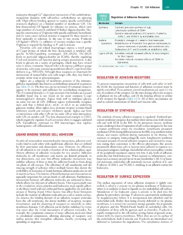
leukocytes through Ca -dependent interactions of the carbohydrate- TABLE
recognition domains with cell-surface carbohydrates on apposing 12.5 Regulation of Adhesion Receptors
cells. High-affinity binding appears to require specific carbohydrate
structures displayed on a limited number of membrane GPs. The Mechanism Example
best-characterized GP ligands for selectins are mucins, which have Synthesis Erythroid precursor synthesis of α 5 β 1
large numbers of clustered, sialylated O-linked oligosaccharides. Site- Lymphocyte synthesis of CD44
specific construction of O-glycans with specific sialylated, fucosylated, Cytokine-induced synthesis of E-selectin, P-selectin,
and (in some cases) sulfated moieties is required for these mucins to ICAM-1, and VCAM-1 by endothelial cells
bind optimally to selectins. In the case of one mucin, P-selectin
GP ligand-1 (PSGL-1), sulfation of tyrosine residues near a specific Surface Proteolytic cleavage of L-selectin from leukocytes
O-glycan is required for binding to P- and L-selectin. expression Redistribution of P-selectin from granule membranes to
Dendritic cells and related macrophages express a novel group plasma membrane of platelets and endothelial cells
of C-type lectins, of which the best characterized is dendritic cell- Endocytosis of P- and E-selectin on endothelial cells
specific ICAM-3-grabbing nonintegrin (DC-SIGN). DC-SIGN Ligand affinity Activation-induced increased affinity of many integrins
binds to particular oligosaccharides on ICAMs, thereby regulating for their ligands
T-cell and dendritic cell function during antigen presentation. It also Activation-induced increased affinity of CD44 for
binds to glycans on a variety of pathogens, which may have critical hyaluronan
roles in innate immunity. Natural killer cells express a different group For abbreviations, see Table 12.1 footnotes.
of proteins, with some containing membrane-distal C-type lectin-like
domains (e.g., NKG2D). Although these receptors are important for
interactions of natural killer cells with target cells, they may bind to
proteins rather than to glycoconjugates. REGULATION OF ADHESION RECEPTORS
Siglecs are a subgroup of membrane proteins of the immuno-
globulin superfamily that bind to carbohydrates instead of to proteins To prevent inappropriate interactions of cells with each other or with
(see Table 12.2). The first two amino-terminal (N-terminal) domains the ECM, the expression and function of adhesion receptors must be
appear to be necessary and sufficient for carbohydrate recognition. tightly controlled. Three primary control mechanisms are used: (1) the
The N-terminal domain is a V-type structure that includes an unusual rate of synthesis of the receptor, (2) the time during which the receptor
disulfide bond that is not found in the more common C-type is displayed on the cell surface, and (3) the binding affinity or avidity
immunoglobulin domains. Siglecs bind well to sialylated glycans of the receptor for ligands (Table 12.5). All of these mechanisms are
on some but not all GPs. Different siglecs preferentially recognize used to control interactions of blood and vascular cells.
sialic acid that is linked α2,6-, α2,8-, or α2,3- to an underlying
galactose residue. Most siglecs have immune receptor tyrosine-based
inhibitory motifs and transmit inhibitory signals. Siglecs can form REGULATION OF SYNTHESIS
cis interactions with other GPs on the same cell or trans interactions
with GPs on another cell. The best-characterized example is CD22, The synthesis of many adhesion receptors is regulated. Erythroid pre-
which negatively regulates B-cell activation when it engages sialylated cursors synthesize integrins that mediate their interactions with stromal
GPs. Sialoadhesin, expressed on BM macrophages, may regulate cells and with ECM in the BM. As the precursors mature, synthesis
hematopoietic cell differentiation. ceases, resulting in loss of expression of cell-surface integrins by the time
a mature erythrocyte enters the circulation. Lymphocyte precursors
synthesize CD44 during differentiation in the BM, stop synthesis before
LIGAND BINDING VERSUS CELL ADHESION release, and resume synthesis during maturation in the thymus. On
exposure to antigens, immunologically naive lymphocytes synthesize
As with all noncovalent macromolecular interactions, adhesion mol- increased amounts of several adhesion receptors and chemokine recep-
ecules bind to each other with equilibrium affinities that are defined tors during their conversion to the effector phenotypes; this process
by their association and dissociation rates. However, the efficiency presumably allows these cells to become more adhesive in response to a
of cell adhesion is not simply a function of the solution-phase equi- subsequent antigenic challenge. Endothelial cells in postcapillary venules
librium affinities of adhesion molecules for one another. Adhesion of the peripheral vasculature express very low, if any, levels of adhesion
molecules in cell membranes and matrix are limited primarily to molecules that bind leukocytes. When exposed to inflammatory cyto-
two dimensions, and even low-affinity molecular interactions may kines such as tumor necrosis factor-α and interleukin-1 (IL-1) or bacte-
stabilize adhesion if there is time for sufficient bonds to form along rial endotoxin, endothelial cells transiently increase synthesis of E- and
the plane of cell contact. The efficiency of cell attachment and the P-selectin, ICAM-1, and VCAM-1, resulting in an adhesive surface for
ensuing strength of adhesion reflect multiple factors that dictate the leukocytes.
probability of formation of bonds between adhesion molecules on cell
or matrix surfaces. The kinetics of bond formation and dissociation are
especially important for certain kinds of cell adhesion. Furthermore, REGULATION OF SURFACE EXPRESSION
interactions between cell adhesion molecules are subjected to force,
which affects the lifetimes of adhesive bonds. This is particularly true The surface expression of some adhesion receptors is tightly con-
in the circulation, where platelets and leukocytes must rapidly adhere trolled. L-selectin is present on the plasma membrane of leukocytes,
to the blood vessel wall and withstand forces applied by the wall shear where it is available to bind to ligands on the endothelial cell surface.
stresses of flowing blood. Other factors that affect bond formation Stimulation of the leukocyte causes L-selectin to be shed into the
include the number of adhesion molecules on a cell or matrix surface, plasma by proteolytic cleavage. P-selectin is constitutively synthesized
the distance the binding domain of an adhesion receptor protrudes by megakaryocytes (where it is incorporated into platelets) and by
from the cell membrane, the lateral mobility of receptors, receptor endothelial cells. Rather than being directly delivered to the plasma
dimerization, and the clustering of receptors on microvilli or other membrane, it is sorted into secretory storage granules: the α granules
membrane domains. Cell adhesion can be further stabilized by events of platelets and the Weibel–Palade bodies of endothelial cells. On
that occur after the initial interactions of adhesion molecules. For stimulation of these cells by agonists such as thrombin, P-selectin is
example, the cytoplasmic domains of many adhesion molecules bind rapidly transported to the cell surface during fusion of granule mem-
to cytoskeletal components, allowing clustering of receptors into branes with the plasma membrane. When they are on the surface of
surface patches that strengthen adhesion, thereby promoting cell the endothelium, both E-selectin and P-selectin are internalized and
spreading or migration. delivered to lysosomes for degradation. The cytoplasmic domain of