
Page 174 - Hematology_ Basic Principles and Practice ( PDFDrive )
P. 174
132 Part II Cellular Basis of Hematology
P-selectin contains signals that direct sorting into secretory granules, Tyrosine kinases have been localized at the interaction zones between
internalization through coated pits of the plasma membrane, and integrins, the cytoskeleton and several adaptor and effector molecules,
movement from endosomes to lysosomes; the latter two signals are and tyrosine phosphorylation of a number of proteins accompanies
probably also present in the cytoplasmic domain of E-selectin. The integrin-mediated cell signaling. Tyrosine phosphorylation initiates
net result of these events is to control the duration of exposure a cascade of signaling events, including the activation of serine/
of E- and P-selectin on the endothelium, where they can mediate threonine kinases, which cause a variety of cellular responses. Ligand
adhesion of leukocytes. Activation of leukocytes also mobilizes a pool binding to integrins also results in generation of lipid second mes-
2+
of β 2 integrins from storage compartments to the plasma membrane, sengers, alkalization of the cytoplasm, and influxes of Ca .
although some of these molecules are also constitutively expressed on
the cell surface. Finally, platelet activation redistributes a portion of
the GPIb–IX–V complexes from ligand-accessible positions on the COOPERATIVE INTERACTIONS BETWEEN SIGNALING
plasma membrane to sequestered, invaginated membrane domains AND ADHESION MOLECULES
known as the surface-connected canalicular system. This process, which
requires interactions of the cytoplasmic domain of GPIb–IX–V with Signaling and adhesion molecules frequently function cooperatively
the cytoskeleton, may serve to downregulate GPIb-mediated adhe- in sequential cascades to enhance the specificity of cell adhesion.
sion of platelets to immobilized vWF. Three examples of how these cooperative interactions facilitate blood
cell responses are described next.
REGULATION OF BINDING AFFINITY
Platelet Adhesion and Aggregation
Regulation of binding affinity is an important control mechanism for
other adhesion receptors. Many integrins are constitutively present on At sites of blood vessel injury in the arterial circuit, platelets
the cell surface but interact poorly with their ligands. Cell activation rapidly tether to and then translocate or roll along the damaged
by a number of agonists induces conformational changes in integrins vessel through reversible interactions of GPIb–IX–V receptors with
so that they effectively recognize their ligands. An example is the immobilized vWF exposed in the subendothelial matrix of injured
α IIb β 3 integrin, which requires platelet stimulation to bind fibrinogen; vessels (Fig. 12.3). These interactions are facilitated by arterial flow,
if this binding affinity were not regulated, circulating platelets would perhaps because of complex effects of high wall shear stresses on the
indiscriminately aggregate in the fibrinogen-rich plasma milieu. The lifetimes of bonds between GPIb and vWF. An important feature
cytoplasmic domains of integrins can exert both positive and negative of this initial reversible adhesive event is that prior activation of the
influences on binding affinity. Binding of specific cytoplasmic proteins platelets is not required. After adhesion, however, the interaction of
to these domains may propagate structural changes to the extracellular immobilized vWF with GPIb receptors triggers intracellular signals
ligand-binding regions of the integrins. Three-dimensional structures that lead to platelet activation. These signals synergize with those
of integrins suggest that the integrin “headpiece” that contains the produced by engagement of the collagen receptor GPVI. Platelet
ligand-binding site faces down toward the membrane in the inactive activation, in turn, increases the affinity of platelet integrins for
conformation and rapidly extends upward in a “switchblade”-like collagen and fibronectin, which stabilizes adhesion. Binding of these
opening motion on activation. Low-affinity ligand binding may ligands transduces signals that propagate further activation responses
stabilize some active conformations of integrins, perhaps explaining such as spreading, secretion of granule contents, and recruitment of
why integrins on unactivated cells will sometimes bind to immobilized, additional platelets through cell–cell contact mediated by binding of
multivalent adhesive proteins but not to the same proteins in solu- fibrinogen to activated α IIb β 3 integrins. This adhesion cascade allows
tion. Cellular activation may also regulate the binding avidities of
CD44, L-selectin, P-selectin, and some integrins through changes in
membrane distribution engineered by interactions of their cytoplasmic
domains with the cytoskeleton or with clathrin-coated pits. Endothelial cells
CELL SIGNALLING THROUGH ADHESION MOLECULES Blood flow Tight
Neutrophil Rolling adhesion Emigration
In addition to their roles in cell–cell and cell–matrix contacts, adhe-
sion molecules may cause cell signaling through indirect or direct
mechanisms. Proteoglycans in the ECM can sequester growth factors
that can be released to bind to surface receptors on nearby cells.
Some chemoattractants bind to proteoglycans on the surface of endo-
thelial cells, where they can activate adherent leukocytes. Binding
of adhesive ligands to cell-surface integrins, GPIb–IX–V, CD44, Selectins Integrins Integrins and
cadherins, CD36, PECAM-1, selectins, ICAM-1 and VCAM-1, PECAM-1
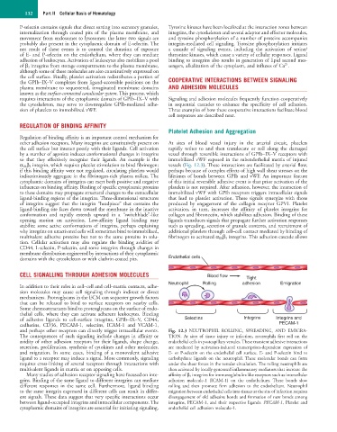
and perhaps other receptors can directly trigger intracellular events. Fig. 12.3 NEUTROPHIL ROLLING, SPREADING, AND EMIGRA-
The consequences of such signaling include changes in affinity or TION. At sites of tissue injury or infection, neutrophils first roll on the
avidity of other adhesion receptors for their ligands, shape change, endothelial cells in postcapillary venules. These transient adhesive interactions
secretion, proliferation, synthesis of cytokines and other molecules, are mediated by activation-induced transcription-dependent expression of
and migration. In some cases, binding of a monovalent adhesive E- or P-selectin on the endothelial cell surface. E- and P-selectin bind to
ligand to a receptor may induce a signal. More commonly, signaling carbohydrate ligands on the neutrophil. These molecular bonds can form
requires cross-linking of several receptors through interactions with under the shear forces in the venular circulation. The rolling neutrophils are
multivalent ligands in matrix or on apposing cells. then activated by locally generated inflammatory mediators that increase the
Many studies of adhesion receptor signaling have focused on inte- affinity of β 2 integrins for immunoglobulin-like receptors such as intercellular
grins. Binding of the same ligand to different integrins can mediate adhesion molecule-1 (ICAM-1) on the endothelium. These bonds slow
different responses in the same cell. Furthermore, ligand binding rolling and then promote firm adhesion to the endothelium. Neutrophil
to the same integrin expressed in different cells can result in differ- migration between endothelial cells into tissues at the site of infection requires
ent signals. These data suggest that very specific interactions occur disengagement of old adhesive bonds and formation of new bonds among
between ligand-occupied integrins and intracellular components. The integrins, PECAM-1, and their respective ligands. PECAM-1, Platelet and
cytoplasmic domains of integrins are essential for initiating signaling. endothelial cell adhesion molecule-1.