
Page 2026 - Hematology_ Basic Principles and Practice ( PDFDrive )
P. 2026
Chapter 119 Transfusion Reactions to Blood and Cell Therapy Products 1797
immediate, or there may be a delay of several hours before the that activate the neutrophils in the lung parenchyma, leading to edema.
symptoms become apparent. Especially for gram-positive bacteria, a Complement and monocyte activation with aggregation of white blood
reaction to infusion of a contaminated unit may not be distinguish- cells also may occur when leukoagglutinins present in the recipient react
able from an FNHTR. Shock in a septic transfusion reaction is with leukocytes contained in the infused donor blood. As a result of the
attributable to endotoxin produced by gram-negative bacteria. Septic leukocyte antigen-antibody reaction, the activated leukocytes express
transfusions differ from acute hemolytic reactions most notably by adhesive molecules on their surface (CD11/CD18), which then permit
the absence of characteristic hemoglobinuria and hemoglobinemia. leukocytes to attach to pulmonary endothelial cells and migrate to the
For a patient who appeared well and suddenly develops rigors, interstitial space between the pulmonary capillaries and the alveolar
fever, and/or shock during an infusion, an infected component epithelium. Once in the interstitial space, neutrophils degranulate and
should be considered. Blood infusion should be stopped the moment through enzymatic digestion produce capillary dehiscence that results
any transfusion reaction is suspected, and appropriate samples should in fluid filling the alveolar sacs. Pulmonary leukostasis with pulmonary
be sent to the blood bank for a DAT, hemolysis check, and bacterial edema thus occurs as a result of microvascular occlusion and capillary
culture. Broad-spectrum antibiotics should be started immediately if leakage. Complement-activated granulocytes also produce oxygen
infusion of contaminated blood is suspected and continued until the radicals that damage pulmonary endothelial cells, resulting in a further
culture results are reported. It is also important to consider a bacteri- increase in pulmonary vascular permeability and additional passage of
ally contaminated blood component when a patient presents with fluid into alveolar spaces. It has been reported that aged blood prod-
signs of bacteremia several hours after a transfusion is completed. ucts may accumulate bioactive lipids and soluble mediators, such as
Gram-positive bacteria, which are the most common bacterial con- CD40L, that hamper the chemokine scavenging ability of erythrocytes
taminants in platelet components, are less likely to cause shock, and as a result of reduction in the expression of the Duffy antigens, and
presentation of signs and symptoms of infection may be delayed by this may represent a second hit in the two-hit model. Rodent model
several hours. systems for TRALI have also been described, and some of these models
Because of the decrease in viral transmission by blood transfusion, suggest a role for platelets in TRALI. Work is ongoing to elucidate the
septic transfusion reactions now account for a significant portion of different mechanisms leading to this syndrome, which may be a final
the transfusion-related infections in the United States. Data from the common pathway from a variety of initiating insults.
Bacterial Contamination of Blood study showed that from 1998 to Because TRALI is hard to distinguish from fluid overload without
2000, the rate of transfusion-transmitted bacteremia was 9.98 per central cardiovascular pressure measurements, it is often not straight-
million single-donor platelets, 10.64 per million pooled platelets, and forward to diagnose. When a patient shows signs of noncardiogenic
0.21 per million RBC units; the rate of fatal reactions was 1.94 per pulmonary edema, the infusion should be immediately stopped, as it
million single-donor platelets, 2.22 per million pooled platelets, and should be with all other reactions. These HLA/neutrophil antigen-
0.13 per million RBC units, respectively. To decrease the likelihood antibody reactions are usually donor-specific and should not recur
of a septic unit of platelets being transfused, the expiration date of with a unit from a different donor.
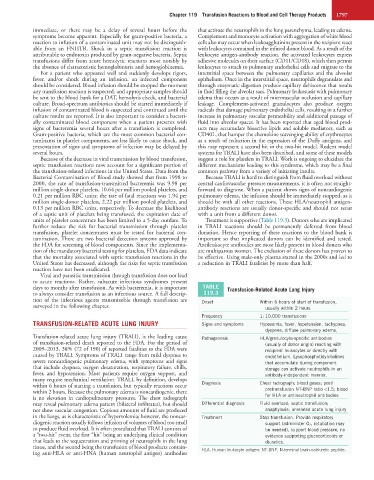
units of platelet concentrate has been limited to a 5-day outdate. To Treatment is supportive (Table 119.3). Donors who are implicated
further reduce the risk for bacterial transmission through platelet in TRALI reactions should be permanently deferred from blood
transfusion, platelet concentrates must be tested for bacterial con- donation. Hence reporting of these reactions to the blood bank is
tamination. There are two bacterial detection systems approved by important so that implicated donors can be identified and tested.
the FDA for screening of blood components. Since the implementa- Antileukocyte antibodies are most likely present in blood donors who
tion of the mandatory bacterial testing for platelets, FDA data indicate are multiparous women. The exclusion of these donors has proven to
that the mortality associated with septic transfusion reactions in the be effective. Using male-only plasma started in the 2000s and led to
United States has decreased, although the risks for septic transfusion a reduction in TRALI fatalities by more than half.
reaction have not been eradicated.
Viral and parasitic transmission through transfusion does not lead
to acute reactions. Rather, subacute infectious syndromes present
days to months after transfusion. As with bacteremia, it is important TABLE Transfusion-Related Acute Lung Injury
to always consider transfusion as an infectious source. A full descrip- 119.3
tion of the infectious agents transmissible through transfusion are Onset Within 6 hours of start of transfusion,
surveyed in the following chapter. usually within 2 hours
Frequency 1 : 10,000 transfusions
TRANSFUSION-RELATED ACUTE LUNG INJURY Signs and symptoms Hypoxemia, fever, hypotension, tachypnea,
dyspnea, diffuse pulmonary edema,
Transfusion-related acute lung injury (TRALI), is the leading cause Pathogenesis HLA/granulocyte-specific antibodies
of transfusion-related death reported to the FDA. For the period of (usually of donor origin) reacting with
2009–2013, 38% (72 of 190) of reported fatalities to the FDA were recipient leukocytes or directly with
caused by TRALI. Symptoms of TRALI range from mild dyspnea to endothelium. Lysophosphotidylcholines
severe noncardiogenic pulmonary edema, with symptoms and signs that accumulate during component
that include dyspnea, oxygen desaturation, respiratory failure, chills, storage can activate neutrophils in an
fever, and hypotension. Most patients require oxygen support, and antibody-independent manner.
many require mechanical ventilation. TRALI, by definition, develops
within 6 hours of starting a transfusion, but typically reactions occur Diagnosis Chest radiograph; blood gases; post/
within 2 hours. Because the pulmonary edema is noncardiogenic, there pretransfusion NT-BNP ratio <1.5; blood
is no elevation in cardiopulmonary pressures. The chest radiograph for HLA or antineutrophil antibodies
may reveal pulmonary edema pattern (bilateral infiltrates), but should Differential diagnosis Fluid overload; septic transfusion;
not show vascular congestion. Copious amounts of fluid are produced anaphylaxis; unrelated acute lung injury
in the lungs, as is characteristic of hypervolemia; however, the noncar- Treatment Stop transfusion. Provide respiratory
diogenic reaction usually follows infusion of volumes of blood too small support (administer O 2 , intubation may
to produce fluid overload. It is often postulated that TRALI consists of be needed), support blood pressure, no
a “two-hit” event, the first “hit” being an underlying clinical condition evidence supporting glucocorticoids or
that leads to the sequestration and priming of neutrophils in the lung diuretics.
tissue, and the second being the transfusion of blood products contain- HLA, Human leukocyte antigen; NT-BNP, N-terminal brain-natriuretic peptide.
ing anti-HLA or anti-HNA (human neutrophil antigen) antibodies