
Page 2027 - Hematology_ Basic Principles and Practice ( PDFDrive )
P. 2027
1798 Part XI Transfusion Medicine
TRANSFUSION-ASSOCIATED CIRCULATORY OVERLOAD
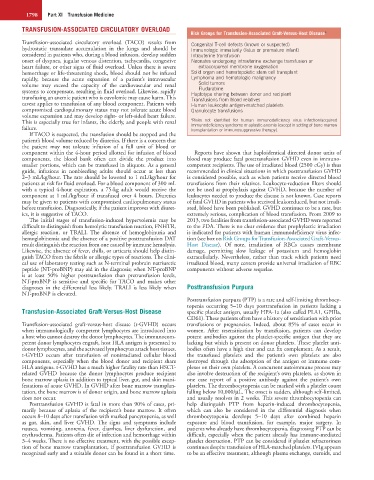
Risk Groups for Transfusion-Associated Graft-Versus-Host Disease a
Transfusion-associated circulatory overload (TACO) results from Congenital T-cell defects (known or suspected)
hydrostatic transudate accumulation in the lungs and should be Immunologic immaturity (fetus or premature infant)
considered in patients who, during a blood infusion, develop sudden Intrauterine transfusion
onset of dyspnea, jugular venous distention, tachycardia, congestive Neonates undergoing intrauterine exchange transfusion or
heart failure, or other signs of fluid overload. Unless there is severe extracorporeal membrane oxygenation
hemorrhage or life-threatening shock, blood should not be infused Solid organ and hematopoietic stem cell transplant
rapidly, because the acute expansion of a patient’s intravascular Lymphoma and hematologic malignancy
volume may exceed the capacity of the cardiovascular and renal Solid tumors
Fludarabine
systems to compensate, resulting in fluid overload. Likewise, rapidly Haplotype sharing between donor and recipient
transfusing an anemic patient who is euvolemic may cause harm. This Transfusions from blood relatives
caveat applies to transfusion of any blood component. Patients with Human leukocyte antigen-matched platelets
compromised cardiopulmonary status may not tolerate acute blood Granulocyte transfusions
volume expansion and may develop right- or left-sided heart failure.
Risks not identified for human immunodeficiency virus infection/acquired
This is especially true for infants, the elderly, and people with renal a immunodeficiency syndrome or aplastic anemia (except in setting of bone marrow
failure. transplantation or immunosuppressive therapy).
If TACO is suspected, the transfusion should be stopped and the
patient’s blood volume reduced by diuretics. If there is a concern that
the patient may not tolerate infusion of a full unit of blood or
component within the 4-hour period allotted for infusion of blood Reports have shown that haploidentical directed donor units of
components, the blood bank often can divide the product into blood may produce fatal posttransfusion GVHD even in immuno-
smaller portions, which can be transfused in aliquots. As a general competent recipients. The use of irradiated blood (2500 cGy) is thus
guide, infusions in nonbleeding adults should occur at less than recommended in clinical situations in which posttransfusion GVHD
2–3 mL/kg/hour. The rate should be lowered to 1 mL/kg/hour for is considered possible, such as when patients receive directed blood
patients at risk for fluid overload. For a blood component of 300 mL transfusions from their relatives. Leukocyte-reduction filters should
with a typical 4-hour expiration, a 75-kg adult would receive the not be used as prophylaxis against GVHD, because the number of
component at 1 mL/kg/hour if transfused over 4 hours. Diuretics leukocytes needed to produce the disease is not known. Case reports
may be given to patients with compromised cardiopulmonary status of fatal GVHD in patients who received leukoreduced, but not irradi-
before transfusion. Diagnostically, if the patient improves with diuret- ated, blood have been published. GVHD continues to be a rare, but
ics, it is suggestive of TACO. extremely serious, complication of blood transfusion. From 2009 to
The initial stages of transfusion-induced hypervolemia may be 2013, two fatalities from transfusion-associated GVHD were reported
difficult to distinguish from hemolytic transfusion reaction, FNHTR, to the FDA. There is no clear evidence that prophylactic irradiation
allergic reaction, or TRALI. The absence of hemoglobinuria and is indicated for patients with human immunodeficiency virus infec-
hemoglobinemia and the absence of a positive posttransfusion DAT tion (see box on Risk Groups for Transfusion-Associated Graft-Versus-
result distinguish the reaction from one caused by immune hemolysis. Host Disease). Of note, irradiation of RBCs causes membrane
Likewise, the absence of fever, chills, or urticaria should help distin- damage, permitting slow leakage of potassium and hemoglobin
guish TACO from the febrile or allergic types of reactions. The clini- extracellularly. Nevertheless, rather than track which patients need
cal use of laboratory testing such as N-terminal probrain natriuretic irradiated blood, many centers provide universal irradiation of RBC
peptide (NT-proBNP) may aid in the diagnosis; when NT-proBNP components without adverse sequelae.
is at least 50% higher posttransfusion than pretransfusion levels,
NT-proBNP is sensitive and specific for TACO and makes other
diagnoses in the differential less likely. TRALI is less likely when Posttransfusion Purpura
NT-proBNP is elevated.
Posttransfusion purpura (PTP) is a rare and self-limiting thrombocy-
topenia occurring 5–10 days posttransfusion in patients lacking a
Transfusion-Associated Graft-Versus-Host Disease specific platelet antigen, usually HPA-1a (also called PLA1, GPIIIa,
CD61). These patients often have a history of sensitization with prior
Transfusion-associated graft-versus-host disease (t-GVHD) occurs transfusions or pregnancies. Indeed, about 85% of cases occur in
when immunologically competent lymphocytes are introduced into women. After resensitization by transfusion, patients can develop
a host who cannot destroy the donor lymphocytes. The immunocom- potent antibodies against the platelet-specific antigen that they are
petent donor lymphocytes engraft, host HLA antigen is presented to lacking but which is present on donor platelets. These platelet anti-
donor lymphocytes, and the activated lymphocytes attack host tissues. bodies often have a high titer and can fix complement. As a result,
t-GVHD occurs after transfusion of nonirradiated cellular blood the transfused platelets and the patient’s own platelets are also
components, especially when the blood donor and recipient share destroyed through the adsorption of the antigen or immune com-
HLA antigens. t-GVHD has a much higher fatality rate than HSCT- plexes on their own platelets. A concurrent autoimmune process may
related GVHD because the donor lymphocytes produce recipient also involve destruction of the recipient’s own platelets, as shown in
bone marrow aplasia in addition to typical liver, gut, and skin mani- one case report of a positive antibody against the patient’s own
festations of acute GVHD. In GVHD after bone marrow transplan- platelets. The thrombocytopenia can be marked with a platelet count
tation, the bone marrow is of donor origin, and bone marrow aplasia falling below 10,000/µL. The onset is sudden, although self-limited,
does not occur. and usually resolves in 2 weeks. This severe thrombocytopenia can
Posttransfusion GVHD is fatal in more than 90% of cases, pri- help distinguish PTP from heparin-induced thrombocytopenia,
marily because of aplasia of the recipient’s bone marrow. It often which can also be considered in the differential diagnosis when
occurs 8–10 days after transfusion with marked pancytopenia, as well thrombocytopenia develops 5–10 days after combined heparin
as gut, skin, and liver GVHD. The signs and symptoms include exposure and blood transfusion, for example, major surgery. In
nausea, vomiting, anorexia, fever, diarrhea, liver dysfunction, and patients who already have thrombocytopenia, diagnosing PTP can be
erythroderma. Patients often die of infection and hemorrhage within difficult, especially when the patient already has immune-mediated
3–4 weeks. There is no effective treatment, with the possible excep- platelet destruction. PTP can be considered if platelet refractoriness
tion of bone marrow transplantation, if posttransfusion GVHD is continues despite transfusion of HLA-matched platelets. IVIg appears
recognized early and a suitable donor can be found in a short time. to be an effective treatment, although plasma exchange, steroids, and