
Page 216 - Hematology_ Basic Principles and Practice ( PDFDrive )
P. 216
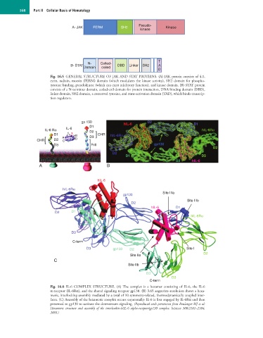
168    Part II  Cellular Basis of Hematology


                                                                  Pseudo-
                                   A- JAK   FERM         SH2                  Kinase
                                                                  kinase


                                                                           T
                                          N-    Coiled-
                                  B- STAT               DBD  Linker  SH2   A
                                        Domain  coiled
                                                                           D
                        Fig. 16.5  GENERAL STRUCTURE OF JAK AND STAT PROTEINS. (A) JAK protein consists of 4.1,
                        ezrin, radixin, moesin (FERM) domain (which modulates the kinase activity), SH2 domain for phospho-
                        tyrosine binding, pseudokinase (which can exert inhibitory function), and kinase domain. (B) STAT protein
                        consists of a N-terminus domain, coiled-coil domain for protein interaction, DNA binding domain (DBD),
                        linker domain, SH2 domain, a conserved tyrosine, and trans-activation domain (TAD), which binds transcrip-
                        tion regulators.


                                       gp 130              hIL-6      D2                   D1
                                           D1
                      IL-6 Rα   IL-6       D2                                                  hIL-6Rα
                          D1    1 3  B        CHR   hIL-6Rα
                              A            D3
                  CHR     D2  C     D
                          D3               Frill                         gp130
                                                                                                    D2
                                                     D2
                                                           D1                                  D3
                                                                                   D2     hIL-6
                   A                               B


                                              hIL-6
                              hIL-6Rα            B
                                                                             Site IIIa
                                                          gp130
                                                                                        Site IIIb
                                                              D2
                                                D    C                 hIL-6       D1
                           D2     D1
                                                A                            D            hIL-6Rα


                                  D3                                   C      B
                                                                          A
                                   C-term
                                         D3           gp130  D2                         Site I
                                                              Site IIa                          D2
                          C
                                                             Site IIb
                                                                     D3
                                                                                 D3
                                                                       C-term
                        Fig.  16.6  IL-6  COMPLEX  STRUCTURE.  (A) The  complex  is  a  hexamer  consisting  of  IL-6,  the  IL-6
                        α-receptor (IL-6Rα), and the shared signaling receptor gp130. (B) 3.65 angström–resolution shows a hexa-
                        meric, interlocking assembly mediated by a total of 10 symmetry-related, thermodynamically coupled inter-
                        faces. (C) Assembly of the hexameric complex occurs sequentially: IL-6 is first engaged by IL-6Rα and then
                        presented to gp130 to activate the downstream signaling. (Reproduced with permission from Boulanger MJ et al:
                        Hexameric  structure  and  assembly  of  the  interleukin-6/IL-6  alpha-receptor/gp130  complex.  Science  300:2101–2104,
                        2003.)