
Page 2255 - Hematology_ Basic Principles and Practice ( PDFDrive )
P. 2255
2002 Part XII Hemostasis and Thrombosis
undergoes a series of posttranslational modifications, including N- or aggregated forms of the protein in heterologous cells with high
and O-linked glycosylation mainly in the B domain (Fig. 135.2) and levels of FVIII expression, their relevance in native FVIII-producing
sulfation of tyrosine residues. Furthermore, when the nascent protein cells (i.e., vascular endothelium and liver sinusoidal endothelial cells)
transits the endoplasmic reticulum (ER), it interacts with ER chap- is unknown. Indeed, under conditions of high-level FVIII expression,
erones, including calreticulin and immunoglobulin binding protein cells can activate a classic unfolded protein response and can succumb
(BiP). Although these interactions limit the transport of malfolded to apoptotic cell death.
The other detail of FVIII trafficking that has attracted attention
is its transit between the ER and Golgi en route to secretion. Efficient
1 7 14 22 23 26 transit through the ER–Golgi boundary appears to require an interac-
5′ 3′ tion with glycans within the B domain of the protein. Absence of
these glycans or lack of specific transport proteins responsible for this
trafficking event can significantly reduce the levels of secreted FVIII.
The Factor VIII Protein Structure
The DNA sequence of the F8 gene predicts a translated single-chain
polypeptide with a molecular weight of 260 kDa consisting of 2351
F8A F8B amino acid residues, including a 19-residue signal peptide. Upon
Fig. 135.1 FACTOR VIII GENE. The factor VIII (FVIII) gene (F8) is translocation to the ER, the signal peptide is cleaved. The remaining
located on the X chromosome at cytogenic band Xq28-qter. The 26 exons polypeptide is 2332 residues long.
span 184 kb of genomic DNA, and there are three open reading frames The FVIII protein consists of three types of domains: the three A
expressed from the locus: the 9 kb FVIII mRNA transcript incorporating all domains, which have a 35% to 40% amino acid sequence homology
26 exons of the gene; the F8A transcript that is transcribed in the opposite to ceruloplasmin and to factor V (FV); a central B domain with no
direction to FVIII and comprises sequences from intron 22; and finally, F8B known homologues; and two C-terminal discoidin-like (discoidin
comprising an initial 5′ exon derived from intron 22 sequence that is spliced being a cell adhesion protein found in slime molds) C domains that
to exons 23 to 26 of the F8 gene. are also 35% to 40% homologous to FV and to ceruloplasmin (Fig.
135.3). There are also three small “a” domains (each between 20 and
40 amino acids) consisting of predominantly acidic residues. The
N-linked glycans sequence of these domains within the protein is NH2-A1-a1-A2-a2-
B-a3-A3-C1-C2-COOH. Tyrosine residues in the a2 and a3 domains,
when sulfated, contribute to the cofactor function of FVIII and enable
its interaction with von Willebrand factor (VWF). As indicated, the
A1 A2 B A3 C1 C2 A and C domains of FVIII have structural similarities to similar A and
C domains in coagulation FV. However, FV does not contain a
domain homologous to the B domain. Given these structural similari-
O-linked glycans ties, it is not surprising that both FVIII and FV function as cofactors
Fig. 135.2 FACTOR VIII GLYCAN MODIFICATION (molecular weight, for serine protease enzymes in the coagulation cascade, FVIII in the
260 kDa; plasma concentration, 100–200 ng/mL; 1 nM). The factor VIII intrinsic tenase complex and FV in the prothrombinase complex.
(FVIII) protein is modified by the addition of multiple N- and O-linked Subsequent proteolysis of the FVIII polypeptide chain in the
glycan chains. The majority of these glycan additions are located in the B Golgi generates a light chain consisting of the A3-C1-C2 domains
domain and appear to play a role in facilitating intracellular trafficking and with a mass of 80 kDa and a heavy chain consisting of the A1 and
secretion of the protein. A2 domains. (see Fig. 135.3).
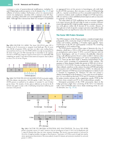
NH2 COOH
A1 A2 B A3 C1 C2
FVIII 372 740 1689
IIa cleavage IIa cleavage IIa cleavage
Me 2+
FVIIIa A1 A2 A3 C1 C2
336 562
APC cleavage
Fig. 135.3 FACTOR VIII PROTEIN ACTIVATION AND INACTIVATION. The factor VIII (FVIII)
protein comprises a series of A and C domains that are homologous to factor V (FV) and ceruloplasmin and
a central B domain that does not show sequence homology. The inactive precursor protein is activated by
thrombin through proteolytic cleavages at three locations. Inactivation of FVIIIa occurs through two mecha-
nisms: spontaneous dissociation of the noncovalently bound A2 domain and proteolysis mediated by activated
protein C (APC).