
Page 2256 - Hematology_ Basic Principles and Practice ( PDFDrive )
P. 2256
Chapter 135 Hemophilia A and B 2003
Circulating FVIII exists as a heterodimer of the NH 2-terminus
heavy chain and the COOH-terminus light chain. The two FVIII
chains are bound to one another at the A1 and A3 domains by
noncovalent bonds that are divalent metal ion dependent. The Enzyme Substrate FXa
FX
FIXa
involved ion is likely copper, which has been found in association
with FVIII.
Cofactor FVIIIa
Storage, Secretion, and Circulation of Factor VIII Gla Gla
Ca 2+ Ca 2+
After synthesis, FVIII is secreted into the circulation, where it forms
a tight noncovalent complex with its multimeric partner VWF (kDa
≈0.2–0.5 nM). Whereas the plasma concentration of FVIII is
100–200 ng/mL (≈1 nM), the concentration of VWF is approxi- Procoagulant phospholipid surface
mately 10 µg/mL (50 nM); thus the molar ratio of FVIII to VWF
in the FVIII-VWF complex is about 1 : 50. The majority of VWF in
plasma is synthesized and secreted by vascular endothelial cells. VWF Fig. 135.4 THE INTRINSIC TENASE COMPLEX. This membrane-
binds to the a3 and C2 regions of FVIII through sequences in the bound complex plays an essential role in the amplification phase of hemostasis.
D′/D3 region of the mature VWF monomer. Just as the cellular Participation of all components of the complex enhances the catalytic effi-
source of FVIII had long been argued, so necessarily has the site at ciency of the FXa-generating process by more than 200,000-fold. Deficiency
which FVIII and VWF first interact. However, the recent finding that or dysfunction of either the enzyme or cofactor involved in the complex
FVIII is expressed by endothelial cells suggests that at least some of results in a marked reduction in catalytic efficiency and the clinical manifesta-
the circulating FVIII may interact with VWF before secretion and tions of hemophilia. FIXa, Factor IXa; FVIIIa, factor VIIIa.
may be co-stored with VWF in Weibel-Palade bodies. In the plasma,
VWF protects FVIII from proteolysis by lipid-binding proteases
including factor Xa (FXa). Without this interaction—for example, in
cases of type 3 von Willebrand disease (VWD; in which VWF is TABLE Genetic Mechanisms Causing Hemophilia in Females
absent) or type 2N VWD (in which mutations occur in the FVIII 135.1
binding region of VWF)—the plasma half-life of FVIII is reduced • F8 or F9 mutation homozygosity
and consequently, the plasma levels of FVIII are low. • F8 or F9 mutation compound heterozygosity
• Extreme skewing of X inactivation process
• X/O karyotype: Turner syndrome
Activation and Coagulant Function of Factor VIII • X/autosome translocation
FVIII plays a critical role in the propagation (amplification) phase of
coagulation. The physiologic activator of FVIII is thrombin, which
proteolytically cleaves FVIII at three sites: Arg372 at the NH 2 - that FVIII clearance may be mainly influenced by its multimeric
terminus of the A2 domain, Arg740 at the NH 2 -terminus of the B partner, VWF, and that a group of lectin and scavenger receptors on
domain, and Arg1689 at the NH 2 -terminus of the A3 domain (see macrophages, and other cells, such as the sinusoidal endothelium in
Fig. 135.3). These cleavages release FVIII from VWF and result in the liver and spleen, may remove FVIII in complex with VWF.
the formation of a noncovalently associated A1-A2-A3/C1/C2 het-
erotrimeric activated FVIII (FVIIIa) molecule. FVIII can also be
activated by FXa and FIXa, although the physiologic contribution of PATHOPHYSIOLOGY OF HEMOPHILIA A
activation by these proteases is less clear.
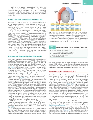
In its activated form, FVIII provides essential cofactor activity in Hemophilia A is a disorder characterized by congenital deficiency of
the intrinsic tenase complex where FIXa is the serine protease and FVIII. Almost all patients with hemophilia A have F8 gene muta-
FX is the substrate (Fig. 135.4). This reaction takes place on a pro- tions. Because F8 is located on the X chromosome, hemophilia A
coagulant phospholipid surface, which in normal hemostasis is likely follows an X-linked inheritance pattern. As a result, most affected
the activated platelet. Participation of FVIIIa in the complex enhances individuals are male. Severe and moderately severe cases of hemophilia
the catalytic efficiency of this reaction about 200,000-fold, and thus A are unusual in females but can result from a number of genetic
severe FVIII deficiency profoundly reduces the rate of FXa generation mechanisms; these are listed in Table 135.1. Approximately 30% to
and renders this reaction biologically futile. The exact details of the 50% of hemophilia A cases are caused by a sporadic mutation and
cofactor role of FVIIIa remain to be elucidated, but it is assumed that occur without a family history of the disorder. The sporadic mutation
it acts as a “scaffold” protein that optimally aligns the enzymatic and is most commonly first evident in a female making her a carrier.
substrate components of the complex on the phospholipid surface. However, as most carriers do not have excessive bleeding symptoms
Indeed, the FIXa and FX interactive regions of FVIII have been the new carrier is unlikely to be diagnosed until later when she has
defined, and the cofactor binds to the phospholipid surface through affected sons of her own or potentially affected grandsons. New
hydrophobic residues in the C2 domain. Of note, the FVIII B female carriers often arise in the context of older paternal age (i.e.,
domain is not required for cofactor function, and to date, it appears older paternal sperm cells). A good example of this was Queen Vic-
that the principal role of this region of the protein is to facilitate toria’s father who was 51 years old at the time that Queen Victoria
trafficking and secretion of the nascent polypeptide. was conceived.
FVIIIa is inactivated through two processes. The predominant The molecular genetic basis of hemophilia A has been extensively
mechanism is through spontaneous dissociation of the A2 domain. characterized over the past 25 years. A comprehensive Internet
The secondary inactivation event is via activated protein C–mediated database of hemophilia A mutations is maintained and updated at
proteolysis at Arg336 and Arg562 in the FVIIIa heavy chain (see http://www.factorviii-db.org.
Fig. 135.3). A significant proportion of hemophilia A cases are the result of a
Our understanding of the clearance of FVIII/FVIIIa is limited. recurrent mutation, an inversion mutation involving sequences in
The low-density lipoprotein (LDL) receptor–related protein and intron 22 of the gene (Fig. 135.5). The mechanism involved in
other members of the LDL receptor family contribute, but this is generating this mutation involves an intrachromosomal recombina-
unlikely to be the complete story. Indeed, evolving evidence indicates tion event in which there is an exchange between the F8A gene in