
Page 2260 - Hematology_ Basic Principles and Practice ( PDFDrive )
P. 2260
Chapter 135 Hemophilia A and B 2007
Hemophilia Carrier Detection and Prenatal Diagnosis
The X-linked recessive nature of hemophilia naturally results in the major- The fourth and most definitive approach to carrier diagnosis is to
ity of affected subjects being males. Females, however, carry and transmit use molecular genetics to identify the causative FVIII or FIX mutation.
the hemophilic trait and can sometimes also express clinical manifesta- Although carrier detection studies originally used analysis of linked poly-
tions of the disease. Although there are rare genetic circumstances that morphisms to track mutant alleles, advances in sequencing technology
can result in moderately severe or severe hemophilia in women (see Table now enable relatively easy access to direct mutation detection. This
135.1), the majority of females with low FVIII or FIX levels have low factor advance has significantly enhanced family counseling for hemophilic
levels because of variable skewing of the random X inactivation process kindred and has also enabled women and their caregivers to prepare
that takes place early during embryonic development. optimally for the delivery of newborns.
There are four ways in which hemophilia carriers can be identified. With current molecular genetic testing strategies, the results of muta-
First, in light of the X-linked transmission of the disease, pedigree analysis tion analyses are available within a few days in urgent circumstances.
will determine the carrier status of some women. Thus all daughters of However, most often results are returned within a few weeks.
hemophilic fathers are obligate carriers, and 50% of the daughters of a If a woman is a carrier, the causative mutation in the FVIII or FIX gene
hemophilia carrier mother will also carry the mutant allele. will be found in 98% of cases. If a mutation is not found, it is possible that
The second mode of identification can be through the manifestation the mutation is deep within an intron or involves a distant transcriptional
of abnormal bleeding caused by a low FVIII or FIX level. Estimates element.
of the percentage of hemophilia carriers who experience excessive Ideally, carrier detection should be performed after puberty but
bleeding (most often demonstrated through menorrhagia) vary but likely before the woman is contemplating starting a family. In many countries,
approximate 20% to 30%. testing for the carrier status of genetic disease is prohibited before
The third mode of carrier detection involves use of the laboratory adolescence so that the girl can participate in discussions of testing
phenotype, in which tests of the intrinsic pathway (PTT) may be abnormal options.
and the plasma levels of FVIII or FIX may be reduced below the normal Prenatal diagnosis of hemophilia should begin with an evaluation of the
range. The etiology for low factor levels in carrier females is multifactorial fetal sex, which can usually be determined through a routine ultrasound
and includes a woman’s blood type and VWF levels in the case of carriers examination. Recent studies suggest that free fetal DNA can be isolated
of FVIII deficiency as well as the ratio of inactivation of the hemophilic from the mother’s blood, with levels increasing toward term. Examination
and normal X chromosomes, a random process that occurs early during of this material for the presence of Y chromosome sequences allows for
embryonic development. If there is markedly skewed inactivation of definitive sex determination. If the fetus is female, no additional studies
the normal X chromosome, the plasma level of FVIII or FIX may be should be performed. If the fetus is male, molecular genetic analysis can
correspondingly reduced, and the carrier female may have a bleeding be used to identify the hemophilic mutation. Fetal DNA can be isolated
disorder. It is important to determine the FVIII or FIX levels in all carrier from chorionic villus samples obtained after 10 weeks of gestation or from
females to provide advice regarding potential bleeding symptoms and the amniocytes obtained by amniocentesis from 12 to 34 weeks. The risk of
risk of bleeding with surgical procedures. However, it should be noted miscarriage with both of these procedures is approximately 1%. If the
that whereas a low plasma level of FVIII or FIX is predictive of a carrier studies are being used for making decisions about therapeutic abortion,
state, the lack of a low factor level does not rule out a carrier state. In the they should be performed as early as possible. Determination of the
case of carriers of FVIII deficiency, a low FVIII to VWF ratio has greater hemophilic status of the fetus will also help plan for delivery, although
predictive power than a low FVIII level on its own, which is only found in consensus about the optimal obstetric management of an affected baby
about 20% of carrier females. is lacking.
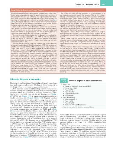
Differential Diagnosis of Hemophilia TABLE
135.4 Differential Diagnosis of a Low Factor VIII Level
The initial clinical suspicion of hemophilia will usually come from
signs and symptoms of excessive bleeding, a family history of a 1. FVIII <10%
bleeding problem, or abnormal coagulation test results. • Severe or moderately severe hemophilia A
There are many causes of mild bleeding manifestations, such as • Severe type 1 VWD
increased bruising and prolonged bleeding after dental and surgical • Type 3 VWD
procedures. These include isolated or combined deficiencies of other • Type 2N VWD
clotting factors (i.e., FXI, FVII, FX, FII, FV deficiency; see Chapter • Acquired hemophilia A
137), VWD (see Chapter 138), or various quantitative or qualitative • Acquired VWD
platelet pathologies (see Chapters 130 to 132). Acquired bleeding 2. FVIII: 10% to 50%
symptoms can be caused by antithrombotic drugs (e.g., antiplatelet • Mild hemophilia A
agents and anticoagulants) or can result from autoantibodies against • Type 1 VWD
clotting factors (e.g., acquired hemophilia A or acquired VWD). • Type 2N VWD
It should be pointed out that a low plasma FVIII level is not • Combined FVIII and FV deficiency
synonymous with a diagnosis of hemophilia A. There are a number FV, Factor V; FVIII, factor VIII; VWD, von Willebrand disease.
of potential diagnoses that can result in low FVIII levels (Table
135.4). Levels of FVIII below 10% are most often the result of
inherited or acquired hemophilia A or a severe form of inherited or
acquired VWD (severe type 1 or type 3 VWD). Determination of FVIII and FV (levels are usually between 5% and 20%) has a preva-
VWF antigen and VWF ristocetin cofactor levels is essential for lence of approximately 1 per million. This rare inherited trait is
diagnosis. Occasionally, levels of FVIII below 10% can be attained caused by recessive mutations in one of two genes involved in the
with type 2N VWD. This diagnosis can be confirmed using FVIII facilitation of protein transport across the ER-Golgi interface: lectin
binding studies or by genotypic analysis of the FVIII binding codons mannose binding protein type 1 or multiple coagulation factor
of VWF (exons 17 to 25, which encode the D′/D3 regions of the deficiency 2.
8
VWF protein). Levels of FVIII between 10% and 50% are also likely Isolated low plasma levels of FIX are almost always caused by
the result of either hemophilia A or VWD (types 1 or one of the type congenital hemophilia B. Interestingly, in contrast to acquired hemo-
2 variants, 2A, 2B, 2M, or 2N). Assessment of VWF levels must be philia A, autoantibody development against FIX is rarely encountered.
undertaken in these cases; if the VWF:RCo/VWF:Ag ratio is below Other situations in which FIX deficiency is found usually involve
0.6, further evaluation of a possible type 2 variant must be pursued. concomitant reductions in the other vitamin K–dependent clotting
Rarely, mild or moderate FVIII deficiency can be co-inherited with factors, such as occurs with vitamin K antagonists, vitamin K defi-
FV deficiency (see Chapter 137). Combined inherited deficiency of ciency, or significant liver disease. A much less common cause of a