
Page 2257 - Hematology_ Basic Principles and Practice ( PDFDrive )
P. 2257
2004 Part XII Hemostasis and Thrombosis
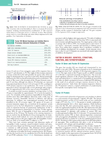
Factor VIII exons 1-22
5′ 3′
F8A F8A F8A
22 14 7 1 1 2 3 4 5 6 7 8
Molecular pathology of hemophilia B
tel cen • No predominant recurring mutation
• Majority of mutations are missense variants
F8B • Some mild phenotypes are due to founder mutations
Fig. 135.5 THE F8 INTRON 22 INVERSION MUTATION. In about Fig. 135.6 THE FACTOR IX GENE (F9). The F9 gene is located on the
45% of patients with severe hemophilia A, there is a recurrent F8 inversion long arm of the X chromosome at cytogenetic band Xq27 centromeric to the
mutation involving intrachromosomal recombination of F8A sequences F8 gene and the common X chromosome fragile site. The gene comprises
within intron 22 of the gene and at a 5′ telomeric location. This mutation 34 kb of genomic DNA arranged in eight exons.
always results in a severe phenotype and almost always originates in the male
germline. Cen, Centromere; tel, telomere.
associated with the highest risk (approximately 75% risk) of inhibitor
development, while the lowest risk (less than 10%) is associated with
TABLE Factor VIII Mutant Genotype and Inhibitor Risk in most missense and splicing mutations. The largest group is those with
135.2 Previously Untreated Hemophilia A Patients an average risk of 20% to 30%; this includes patients with intron 22
Multidomain deletions ≈75% and intron 1 inversions, nonsense and insertion or deletion muta-
Light chain nonsense mutations 30%–40% tions. Even within these categories there is significant variability in
risk of inhibitor development; for example, certain missense muta-
Intron 22 inversion 20%–25% tions in the C2 and C3 domains are associated with much higher risk
Single domain deletions 15%–25% of inhibitor development. 4
Small non-A run insertions/deletions 15%–20%
Heavy chain nonsense mutations 10%–20% FACTOR IX BIOLOGY: GENETICS, STRUCTURE,
Factor VIII missense mutations <10% FUNCTION, AND PATHOPHYSIOLOGY
Small A run insertions/deletions <5% Factor IX Gene and Factor IX Expression
Splicing mutations <5%
The gene that encodes FIX was cloned and characterized by two
groups in the early 1980s. The gene is located on the X chromosome
intron 22 and one of two extragenic copies of this sequence that are at cytogenetic band Xq27 and spans 34 kb of genomic sequence (Fig.
located 5′ and telomeric to F8. The origin of this recurrent mutation 135.6). The eight exons of the F9 gene encode an mRNA transcript
is almost exclusively in the male germline (in sperm cells), where the of 1.4 kb that is expressed exclusively in hepatocytes. The transcrip-
single X chromosome has no partner to pair with, thereby potentially tional regulatory elements of the F9 gene have been well character-
facilitating the F8A-mediated intrachromosomal recombination. The ized, and indeed there is a variant form of hemophilia B, hemophilia
result of this event is the inversion of the F8 gene between exons 1 B Leyden, in which a postpubertal rescue of inherited FIX deficiency
and 22 and thus, although two distinct F8 transcripts are expressed is the result of an activated androgen response element in the F9
5
from separate promoters (exons 1–22 and exons 23–26), a contiguous proximal promoter. Aside from this hormone-responsive element,
FVIII protein sequence is not synthesized. The intron 22 inversion the F9 promoter contains a typical combination of predominantly
mutation is always associated with a severe phenotype and is respon- liver-specific transcription factor binding sites.
sible for approximately 45% of the cases of severe hemophilia A.
A second recurrent inversion mutation involving intron 1 of the
F8 gene is responsible for approximately 2% of severe hemophilia A Factor IX Protein
cases. The remainder of the mutations responsible for hemophilia A
involves more than 2000 different alterations with a wide array of FIX is synthesized exclusively in hepatocytes and circulates in plasma
missense, nonsense, frameshift, insertion or deletion, and splicing at a concentration of approximately 5 µg/mL (90 nM). The mature
mutations. In addition, two transcriptional mutations have been circulating protein consists of 416 amino acids and has a molecular
identified in the F8 promoter region. All regions of the gene are weight of 57 kDa (Fig. 135.7). The protein is initially synthesized as
potential mutation targets, but, as elsewhere in the genome, certain a prepropolypeptide with a short signal sequence, which facilitates
sequences such as the CpG dinucleotide in arginine codons are more entry into the ER, and a propolypeptide sequence, which interacts
prone to mutation because of spontaneous methylation of the 5′ with the γ-glutamylcarboxylase in the ER. The interaction with this
cytosine and spontaneous deamination to thymine. To date approxi- microsomal enzyme catalyzes the posttranslational conversion of the
mately 98% of cases of hemophilia A have been associated with amino-terminal 12 glutamic acid residues of mature FIX into
mutations of the F8 locus. The location of the missing mutations is γ-carboxyglutamyl residues (the GLA domain). This posttranslational
not yet resolved, but the likelihood of locus heterogeneity for hemo- modification enables FIX to form calcium-dependent interactions
philia A seems small. Indeed, the recent identification of mutations with procoagulant phospholipid membranes. A second posttransla-
deep within introns of the gene suggests that all hemophilia A muta- tional modification at residue 64 in the first epidermal growth factor
tions are likely to be found within or adjacent to F8. (EGF)–like domain of the mature polypeptide involves β-hydroxylation
There have been extensive genotype and phenotype studies of of asparagine. This modification also plays a role in facilitating
hemophilia A with, in general, a good correlation between null muta- interactions with calcium and phospholipid membranes.
tions and severe disease and between nonnull missense mutations and The structure of FIX is homologous to that of other vitamin K–
a moderate or mild phenotype. The phenotype remains generally dependent coagulation proteins. The domain structure includes an
consistent with a specific mutation both within and among families. amino-terminal GLA sequence followed by two EGF-like domains,
In addition to its utility for family counseling issues, the F8 geno- an activation peptide and the carboxyl-terminus catalytic domain,
type has also been found to be a predictive factor for the development which is similar to the organization of procoagulant factor VII (FVII)
of anti-FVIII antibodies (Table 135.2). Multidomain deletions are and FX and to the anticoagulant protein C.