
Page 2259 - Hematology_ Basic Principles and Practice ( PDFDrive )
P. 2259
2006 Part XII Hemostasis and Thrombosis
TABLE
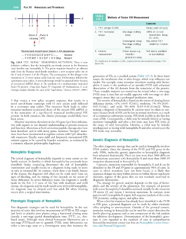
5′ 3′ 135.3 Methods of Factor VIII Measurement
Comments
1 2 3 4 5 6 7 8
1. FVIII antigen ELISA Rarely used clinically
2. FVIII functional One-stage clotting >98% of clinical
Intron 3 Exon 4 assays assay laboratories. Assay
ctcaaag ATC CV <10%.
Chromogenic assay May detect FVIII
g Two-stage clotting instability mutants
assay
Normal and mutant exon 3/exon 4 AA sequences 3. Indirect Global assays e.g., Not readily available
.....Y V D G D Q C E S N P C L..... Normal measurement thrombin or standardized
.....Y V E M E I S V S P I H V Stop Mutant generation assay
CV, Coefficient of variation; ELISA, enzyme-linked immunosorbent assay; FVIII,
Fig. 135.9 THE “ROYAL” HEMOPHILIA MUTATION. There is now factor VIII.
definitive evidence that the hemophilia previously present in the European
royal families was hemophilia B. The point mutation found in an affected
male from the Russian royal family introduces a new acceptor splice site at
the 3′ end of intron 3 of the F9 gene. The consequence of this change is the generation of FXa in a purified system (Table 135.3). In these latter
translation of 11 novel amino acids from the exon 3/4 boundary followed by assays, the incubation time is often longer, which may influence test
a premature stop codon. A severe phenotype would be expected either because results. For example, some missense mutations causing mild hemo-
of accelerated mRNA decay or the synthesis of a nonfunctional truncated philia A result in the synthesis of an unstable FVIII with enhanced
factor IX protein. (Data from Rogaev EI, Grigorenko AP, Faskhutdinova G, et al: dissociation of the A2 domain from the remainder of the protein.
Genotype analysis identifies the cause of the “royal disease.” Science 326:817, 2009.)
These unstable mutants can sometimes be missed when a one-stage
FVIII assay is used but are readily apparent with two-stage or chro-
mogenic assays that use a prolonged incubation time.
3 that creates a new splice acceptor sequence that results in a The severity of hemophilia is based on the extent of clotting factor
novel out-of-frame transcript with 11 new amino acids followed deficiency (severe, <1% [<0.01 IU/mL]; moderate, 1%–5% [0.01–
by a premature stop codon. This mutation likely leads to either 0.05 IU/mL]; and mild, 5%–40% [0.05–0.40 IU/mL]). When
nonsense-mediated accelerated decay of the mutant FIX mRNA or making a diagnosis of hemophilia B, caution must be taken in neo-
to the translation of a significantly truncated nonfunctional FIX nates because levels of FIX can be low in normal newborns as a result
protein. In both instances, the clinical phenotype would likely have of an immature carboxylase system. FIX levels double in the first few
been severe. years of life. Consequently, a child may be initially labeled as having
Missense mutations elsewhere in the FIX gene have been informa- moderate hemophilia and after a few years may have FIX levels in
tive with regards to our detailed understanding of protein structure the range of mild hemophilia. Likewise, a child may be initially
and function. Mutations at both activation peptide cleavage sites have misdiagnosed as having mild hemophilia B and after several years the
been described, and as with many genes, recurrent “hotspot” muta- FIX levels may normalize.
tions have been documented at arginine codons with CpG dinucleo-
tide sequences. Finally, some mild and frequently encountered FIX
variants appear to be caused by founder mutations, as evidenced by Genetic Diagnosis of Hemophilia
a common adjacent polymorphic haplotype.
The other diagnostic strategy that can be used in hemophilia involves
DNA analysis. Since the cloning of the FVIII and FIX genes in the
Hemophilia Diagnosis early 1980s, molecular genetic approaches to hemophilia diagnosis
have advanced dramatically. There are now more than 2000 different
The initial diagnosis of hemophilia depends to some extent on the F8 mutations associated with hemophilia A and more than 1000 F9
family context. In families in which hemophilia has previously been mutations documented in hemophilia B.
identified, family counseling determines the risk of hemophilia Currently, mutations responsible for hemophilia A and B can be
transmission and usually results in a diagnosis being made in utero identified in the F8 and F9 genes in approximately 98% of cases. In
or early in neonatal life. In contrast, when there is no family history cases in which mutations have not been found, it is likely that
of the disease, the diagnosis will often not be made until there are sequence changes are deep within introns or within distant regulatory
signs of bleeding, and the timing of this depends on the extent of elements, regions of the genes that are not routinely examined in
factor deficiency. In severe deficiency states, the diagnosis is usually diagnostic laboratories.
made in the first 1–2 years of life, but with moderate and mild The strategy for genetic analysis depends on the type of hemo-
disease, the diagnosis may be made much later; with mild hemophilia, philia and the severity of the phenotype. For example, all patients
the diagnosis may be delayed until late adult life when bleeding with severe hemophilia A should be screened initially for the recurrent
occurs after a surgical intervention. F8 intron 22 and intron 1 inversion mutations. In contrast, most
patients with hemophilia B require full-sequence analysis of the entire
FIX promoter, coding region, and splice sites.
Phenotypic Diagnosis of Hemophilia When a familial mutation has already been identified in the FVIII
or FIX gene, a prenatal diagnosis can be made by either chorionic
Two diagnostic strategies can be used for hemophilia. In the vast villus sampling or amniocentesis. Outside of the prenatal context,
majority of cases, this involves measurement of FVIII or FIX coagu- mutation testing in hemophilia can be used for carrier detection for
lant levels in platelet poor plasma using a functional clotting assay family planning purposes and as one component of the risk analysis
(usually a one-stage partial thromboplastin time [PTT], i.e., clot- for inhibitor development. Determination of the hemophilic geno-
based assay). Although most clinical hemostasis laboratories use a type is now regarded as the standard of care in comprehensive
one-stage PTT-based test to quantify FVIII, some laboratories use hemophilia treatment centers (see box on Hemophilia Carrier Detec-
either a two-stage assay or a chromogenic assay that measures the tion and Prenatal Diagnosis).