
Page 2326 - Hematology_ Basic Principles and Practice ( PDFDrive )
P. 2326
2068 Part XII Hemostasis and Thrombosis
may also complicate other types of sepsis-induced DIC and may
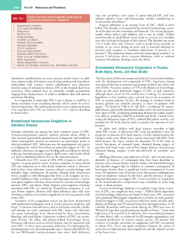
Clinical Conditions Most Frequently Complicated by
BOX 139.1 Disseminated Intravascular Coagulation enhance platelet–vessel wall interactions, thereby contributing to
microvascular thrombosis. 14
• Sepsis/severe infection Purpura fulminans is an extreme form of DIC, which is often
• Trauma/burn/heatstroke lethal. This disorder is characterized by extensive hemorrhagic necro-
• Malignancy sis of the skin over the extremities and buttocks. The disease predomi-
Solid tumors nantly affects infants and children and is rare in adults. Diffuse
Acute leukemia microthrombi in small blood vessels leads to necrosis and vasculitis
• Obstetric conditions may also be found in biopsies of skin lesions. The disorder can occur
Amniotic fluid embolism 2 to 4 weeks after mild infection, such as scarlet fever, varicella, or
Abruptio placentae rubella, or can occur during an acute viral or bacterial infection in
HELLP syndrome
• Vascular abnormalities patients with acquired or hereditary deficiencies of protein C or
Kasabach-Merrit Syndrome protein S. The syndrome mimics neonatal homozygous protein C or
Other vascular malformations protein S deficiency where purpura fulminans, with or without
Aortic aneurysms extensive thrombosis, develops soon after birth.
• Severe allergic/toxic reactions
• Severe immunologic reactions (e.g., transfusion reaction)
Disseminated Intravascular Coagulation in Trauma,
Brain Injury, Burns, and Heat Stroke
thrombotic manifestations are more common in the former. In addi- The time interval between trauma and medical intervention correlates
tion, patients with solid tumors may develop nonbacterial thrombotic with the development and magnitude of DIC. Experience during
endocarditis with systemic arterial embolization and infarction. wars proved that fast evacuation and prompt medical care reduce the
Another cause of subacute to chronic DIC is the retained dead fetus risk of DIC. Extensive exposure of TF to the blood and hemorrhagic
syndrome. These patients have an extremely variable presentation shock are the most immediate triggers of DIC in such instances,
ranging from asymptomatic to mild or moderate skin and mucous although direct proof of this mechanism is lacking. An alternative
membrane bleeding. hypothesis is that cytokines play a pivotal role in the occurrence of
It is important to stress that DIC is not a disease in itself but is DIC in trauma patients. In fact, the changes in cytokine levels in
always secondary to an underlying disorder, which causes the activa- trauma patients are virtually identical to those in patients with
15
tion of coagulation. The underlying disorders most commonly known sepsis. The levels of TNF-α, IL-1β, PAI-1, circulating TF, neutro-
to be associated with DIC are listed in Box 139.1 and are described phil elastase, and soluble thrombomodulin can be elevated in patients
in detail below. with signs of DIC, predicting multiorgan dysfunction (adult respira-
tory distress syndrome [ARDS] included) and death. Careful moni-
Disseminated Intravascular Coagulation in toring for laboratory signs of DIC, reduced fibrinolytic activity, and
perhaps low AT levels may be useful to predict the outcome of such
Infectious Disease patients.
In adults and children with head injuries, mortality is high
Systemic infections are among the most common causes of DIC. when DIC occurs. A laboratory DIC score has predictive value for
Immunocompromised patients, asplenic patients whose ability to prognosis in patients with head injuries, thereby supplementing the
clear bacteria (particularly pneumococci) is impaired, and newborns Glasgow coma score. Brain injury can be associated with DIC, most
whose anticoagulant systems are immature are particularly prone to likely because the injury exposes the abundant TF of the brain to
infection-induced DIC. Infections may be superimposed on trauma blood. Specimens of contused brain, obtained during surgery in
or malignancies, which themselves are potential triggers of DIC. In patients with head injury, and of liver, lungs, kidneys, and pancreas
addition, infections can aggravate bleeding and thrombosis by directly obtained during autopsy, reveal microthrombi in arterioles and
inducing thrombocytopenia, hepatic dysfunction, and shock, which venules.
can lead to diminished blood flow in the microcirculation. Bleeding, laboratory tests indicative of DIC, and vascular micro-
Clinically overt DIC occurs in 30%–50% of patients with gram- thrombi in biopsies of undamaged skin have been described in
negative or gram-positive sepsis. Extreme examples of sepsis-related patients with extensive burns. Kinetic studies with labeled fibrinogen
DIC are streptococcus A toxic shock syndrome, which is character- and platelets suggest that in addition to systemic consumption of
ized by deep tissue infection, vascular collapse, vascular leakage, and hemostatic factors, there is significant local consumption in burned
multiple organ dysfunction. M protein released from streptococci areas. TF exposed at sites of burned tissue, the systemic inflammatory
forms complexes with fibrinogen that bind to β 2 integrins on neu- response syndrome induced by the burn, and the presence of super-
trophils leading to their activation, and meningococcemia, a fulmi- imposed infections can trigger DIC. Local activation of coagulation
nant gram-negative infection characterized by extensive hemorrhagic in the bronchoalveolar compartment may contribute to acute lung
necrosis, DIC, and shock. More frequent gram-negative infections injury in these patients.
associated with DIC are caused by Pseudomonas aeruginosa, E. coli, A severe hemorrhagic diathesis and multiple organ failure indica-
16
and Proteus vulgaris. Patients with these infections may only have tive of DIC can complicate heat stroke. Diffuse fibrin deposition
laboratory evidence of activated coagulation or they may present with and hemorrhagic infarctions are found in fatal cases. DIC associated
severe DIC. with profound fibrin(ogen) degradation is evident in such patients.
Activation of the coagulation system has also been documented Potential triggers of DIC in patients with heat stroke include endo-
with nonbacterial pathogens, such as viruses, protozoa (malaria), and thelial cell damage and TF released from heat-damaged tissues. In 18
13
fungi. Common viral infections, such as influenza, varicella, rubella, critically ill patients with heat stroke during the 2003 heat wave in
16
and rubeola, are rarely associated with DIC. Some viral infections Western Europe that caused numerous deaths in France, there were
can cause hemorrhagic fever characterized by fever, hypotension, high levels of IL-6 and IL-8. In addition, there was marked activation
bleeding, and renal failure. Laboratory evidence of DIC can accom- of white blood cells, as evidenced by β2-integrin upregulation and
pany Korean, rift valley, and dengue-related hemorrhagic fevers. increased production of reactive oxygen species. All patients had
Protozoan infections, such as cerebral malaria, may be associated with evidence of systemic activation of coagulation and DIC was present
overt DIC. In these cases, secondary deficiency of a disintegrin-like in about 35%. There was good correlation between the extent of
metalloprotease with thrombospondin type 1 repeats (ADAMTS13), activation of inflammation and coagulation and the clinical severity
the von Willebrand cleaving protease, may occur. Such deficiency of the heat stroke.