
Page 2386 - Hematology_ Basic Principles and Practice ( PDFDrive )
P. 2386
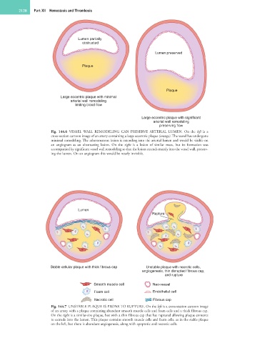
2128   Part XII  Hemostasis and Thrombosis


                                     Lumen partially
                                       obstructed
                                                                        Lumen preserved


                                       Plaque


                                                                             Plaque
                             Large eccentric plaque with minimal
                                 arterial wall remodeling
                                   limiting blood flow


                                                                  Large eccentric plaque with significant
                                                                       arterial wall remodeling
                                                                          preserving flow
                        Fig.  144.6  VESSEL  WALL  REMODELING  CAN  PRESERVE  ARTERIAL  LUMEN.  On  the  left  is  a
                        cross-section cartoon image of an artery containing a large eccentric plaque (orange). The vessel has undergone
                        minimal remodeling. The atheromatous lesion is extending into the arterial lumen and would be visible on
                        an  angiogram  as  an  obstructing  lesion.  On  the  right  is  a  lesion  of  similar  mass,  but  its  formation  was
                        accompanied by significant vessel wall remodeling so that the lesion extends mainly into the vessel wall, preserv-
                        ing the lumen. On an angiogram this would be nearly invisible.


                                     Lumen
                                                                       Rupture


                        Stable cellular plaque with thick fibrous cap  Unstable plaque with necrotic cells,
                                                                  angiogenesis, thin disrupted fibrous cap,
                                                                             and rupture
                                            Smooth muscle cell         Neo-vessel

                                            Foam cell                  Endothelial cell
                                            Necrotic cell              Fibrous cap
                        Fig. 144.7  UNSTABLE PLAQUE IS PRONE TO RUPTURE. On the left is a cross-section cartoon image
                        of an artery with a plaque containing abundant smooth muscle cells and foam cells and a thick fibrous cap.
                        On the right is a similar-size plaque, but with a thin fibrous cap that has ruptured allowing plaque contents
                        to extrude into the lumen. This plaque contains smooth muscle cells and foam cells, as in the stable plaque
                        on the left, but there is abundant angiogenesis, along with apoptotic and necrotic cells.