
Page 256 - Hematology_ Basic Principles and Practice ( PDFDrive )
P. 256
208 Part III Immunologic Basis of Hematology
postcapillary venules, and upon antigenic recognition, remain in the spleen is divided into two functionally and morphologically distinct
node to proliferate and differentiate. If the naive T cells do not compartments, the white pulp and the red pulp (Fig. 19.6). The
encounter antigens they recognize, the cells drain by means of lym- white pulp is composed mainly of lymphoid cells and is the site of
phatic fluid back to the blood and repeat their route in a different antigenic stimulation of B and T cells. The red pulp consists mainly
LN until they become resident lymphocytes or undergo anergy- of myeloid cells, including macrophages that ingest opsonized anti-
mediated cell death (refer to Chapter 21 for further information on gens and damaged erythrocytes from the systemic circulation. The
T-cell immunity). red pulp functions also as a site of extramedullary hematopoiesis early
Egress of lymphocytes from LNs and from the thymus is regulated in fetal life and is a storage site for iron, erythrocytes, and platelets.
by a specialized lipid produced in lymphoid tissue and is known as Extramedullary hematopoiesis in the spleen may also occur postna-
sphingosine-1-phosphate (S1P). Lymphocytes express S1P receptor-1 tally in patients whose BM is incapable of producing adequate
(S1P 1) receptors that facilitate their egress from tissues into blood. numbers of mature blood cells.
Novel immunosuppressive therapeutics antagonizing S1P are being In many mammalian species, including humans, splenic blood
developed; these S1P antagonists reduce release of lymphocytes from flows through a unique vascular circulation that ensures the interpos-
lymphoid tissues into blood. ing of blood (and therefore blood-borne antigens) with the lymphoid
areas of the white pulp. This has been best characterized in the
SECONDARY LYMPHOID TISSUE: COMMON AND murine model. In this model, the splenic white pulp consists of three
compartments—the periarteriolar lymphoid sheath (PALS), follicles,
UNIQUE ANATOMY AND FUNCTIONS and the marginal zone—that interact with blood through an open
sinusoidal arterial network. The PALS is the spleen’s T-cell zone and
In addition to LNs, the spleen is an important site for B-cell develop- is found surrounding the central artery. Follicles in the spleen are
ment and for antigen presentation and stimulation of the adaptive found adjacent to the PALS and are capable of generating primary
immune system. 26–28 Lacking afferent lymphatics, the spleen serves to and secondary follicles with GCs as in LNs. The marginal zone,
sample blood, rather than lymphatic fluid for foreign antigens. The composed of subsets of B cells and macrophages, surrounds the
Capsule
Trabeculum
White pulp
A
Venous sinus
Red pulp
B
T-cell zone (PALS)
B-cell follicles
Marginal sinus
Marginal zone
Follicular arterioles
Central arteriole
Trabecular artery
D
C
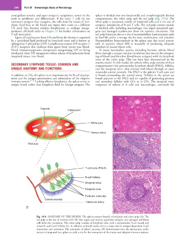
Fig. 19.6 ANATOMY OF THE SPLEEN. The spleen consists broadly of red pulp and white pulp (A). The
red pulp is the site of myeloid cells (B) that ingest and remove opsonized antigens and damaged red blood
cells from the circulation. The white pulp consists of lymphoid cells, with a periarteriolar T-cell sheath and
mixed B- and T-cell follicles (C). In addition to lymph nodes, this is a major site for antigen-dependent B-cell
maturation and activation. The schematic of splenic anatomy (D) demonstrates how the microscopic archi-
tecture is integrated into spleen to yield a site for the interaction of the innate and adaptive immune systems.