
Page 254 - Hematology_ Basic Principles and Practice ( PDFDrive )
P. 254
206 Part III Immunologic Basis of Hematology
naive B cells that have yet to encounter antigen recognized by their
Arterial
surface BCR/Ig complex. Recognition of an antigen by the BCR/Ig
Macrophage leads to B cell activation. A complex set of interactions between the
B cell, T cell, and APCs leads to the activation and proliferation of
B cells carrying an antigen-specific BCR. A fraction of these activated
proliferating B cells form GCs, which are surrounded by a mantle
zone of naive B cells, which together comprise a secondary follicle
(Fig. 19.5).
Germinal centers are classically divided into two compartments,
Dendritic denoted as the dark and light zone based on their appearance under
cell light microscopy (see Fig. 19.5, inset). Dark zones are located adjacent
to the T-cell areas, and contain a high density of proliferating B cells
termed centroblasts, which are large cells with a high nuclear:cytoplasmic
ratio, and do not express surface BCR/Ig. The light zone has a lower
cellular density secondary to the presence of an extensive loose
24
network of follicular DCs, and this imparts the “light” appearance.
Venous Lymphatic B cells in the light zone are termed centrocytes, which in contrast to
the centroblasts, are small B cells expressing surface BCR/Ig. Some
of these cell types, such as centrocytes and centroblasts, are discussed
in Chapter 73 in the context of lymphoid malignancies.
Within the germinal center, the series of sequential shuttling and
reentry of B cells into the dark and light zones is termed the GC
reaction, or the B-cell selection process. The cyclic reentry model
proposes that centroblasts in the dark zone undergo cell division and
SHM of variable light-chain genes mediated by AID. Next, they
reexpress BCR/sIg and exit the cell cycle, migrating into the light
zone to interact with antigen-presenting follicular DCs and TFH
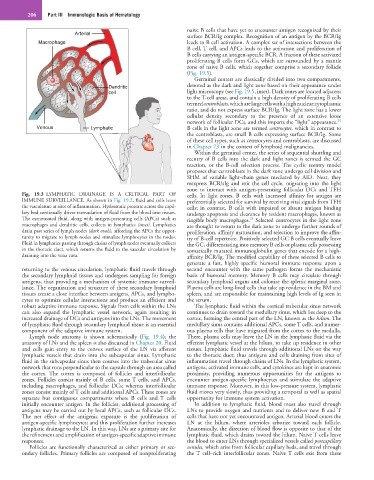
Fig. 19.3 LYMPHATIC DRAINAGE IS A CRITICAL PART OF cells. In light zones, B cells with increased affinity for antigen are
IMMUNE SURVEILLANCE. As shown in Fig. 19.2, fluid and cells leave preferentially selected for survival by receiving vital signals from TFH
the vasculature at sites of inflammation. Hydrostatic pressure across the capil- cells; in contrast, B cells with impaired or absent antigen binding
lary bed continually drives transudation of fluid from the blood into tissues. undergo apoptosis and clearance by resident macrophages, known as
The extravasated fluid, along with antigen-presenting cells (APCs) such as tingible body macrophages. Selected centrocytes in the light zone
25
macrophages and dendritic cells, collects in lymphatics (inset). Lymphatics are thought to return to the dark zone to undergo further rounds of
drain past series of lymph nodes (dark ovals), affording the APCs the oppor- proliferation, affinity maturation, and selection to improve the affin-
tunity to migrate to lymph nodes and stimulate lymphocytes in the nodes. ity of B-cell repertoire. Positively selected GC B cells eventually leave
Fluid in lymphatics passing through chains of lymph nodes eventually collects the GC, differentiating into memory B cells or plasma cells possessing
in the thoracic duct, which returns the fluid to the vascular circulation by somatically mutated immunoglobulin genes that encode for a high-
draining into the vena cava. affinity BCR/Ig. The modified capability of these selected B cells to
generate a fast, highly specific humoral immune response upon a
returning to the venous circulation, lymphatic fluid travels through second encounter with the same pathogen forms the mechanistic
the secondary lymphoid tissues and undergoes sampling for foreign basis of humoral memory. Memory B cells may circulate through
antigens, thus providing a mechanism of systemic immune surveil- secondary lymphoid organs and colonize the splenic marginal zone.
lance. The organization and structure of these secondary lymphoid Plasma cells are long-lived cells that take up residence in the BM and
tissues create a close interface between antigens, APCs, and lympho- spleen, and are responsible for maintaining high levels of Ig seen in
cytes to optimize cellular interactions and produce an efficient and the serum.
robust adaptive immune response. Signals from cells within the LNs The lymphatic fluid within the cortical trabecular sinus network
can also expand the lymphatic vessel network, again resulting in continues to drain toward the medullary sinus, which lies deep to the
increased drainage of DCs and antigens into the LNs. The movement cortex, forming the central part of the LN, known as the hilum. The
of lymphatic fluid through secondary lymphoid tissue is an essential medullary sinus contains additional APCs, some T cells, and numer-
component of the adaptive immune system. ous plasma cells that have migrated from the cortex to the medulla.
Lymph node anatomy is shown schematically (Fig. 19.4); the There, plasma cells may leave the LN in the lymphatic fluid via the
anatomy of LNs and the spleen is also discussed in Chapter 20. Fluid efferent lymphatic vessel at the hilum, to take up residence in other
and cells gain entry to the convex surface of the LN via afferent tissues. Lymphatic fluid travels through additional LNs on the way
lymphatic vessels that drain into the subcapsular sinus. Lymphatic to the thoracic duct; thus antigens and cells draining from sites of
fluid in the subcapsular sinus then courses into the trabecular sinus inflammation travel through chains of LNs. In the lymphatic system,
network that runs perpendicular to the capsule through an area called antigens, activated immune cells, and cytokines are kept in anatomic
the cortex. The cortex is composed of follicles and interfollicular proximity, providing numerous opportunities for the antigens to
zones. Follicles consist mainly of B cells, some T cells, and APCs, encounter antigen-specific lymphocytes and stimulate the adaptive
including macrophages, and follicular DCs; whereas interfollicular immune response. Moreover, in this low-pressure system, lymphatic
zones consist mainly of T cells and additional APCs. These zones are fluid moves very slowly, thus providing a temporal as well as spatial
separate but contiguous compartments where B cells and T cells opportunity for immune system activation.
initially encounter antigen. In the follicles, additional processing of In addition to lymphatic fluid, blood must also travel through
antigens may be carried out by local APCs, such as follicular DCs. LNs to provide oxygen and nutrients and to deliver new B and T
The net effect of the antigenic exposure is the proliferation of cells that have not yet encountered antigen. Arterial blood enters the
antigen-specific lymphocytes; and this proliferation further increases LN at the hilum, where arterioles arborize toward each follicle.
lymphatic drainage to the LN. In this way, LNs are a primary site for Anatomically, the direction of blood flow is opposite to that of the
the refinement and amplification of antigen-specific adaptive immune lymphatic fluid, which drains toward the hilum. Naive T cells leave
responses. the blood to enter LNs through specialized vessels called postcapillary
Follicles are functionally characterized as either primary or sec- venules, which arise from follicular capillary beds, and travel through
ondary follicles. Primary follicles are composed of nonproliferating the T cell–rich interfollicular zones. Naive T cells exit from these