Page 255 - Hematology_ Basic Principles and Practice ( PDFDrive )
P. 255

Chapter 19  Overview and Compartmentalization of the Immune System  207


                                                           Artery Vein
                                     Efferent
                                     lymphatic
                                                                      Medulla


                              Primary
                              follicles
                                                                                            High
                                                         Medullary sinus                    endothelial
                                                                                            venule
                           Secondary
                              follicles
                                Mantle                                                     Cortex
                                 zone


                                     Germinal
                                       center
                                            T-cell
                                            zone
                                                        Afferent lymphatics
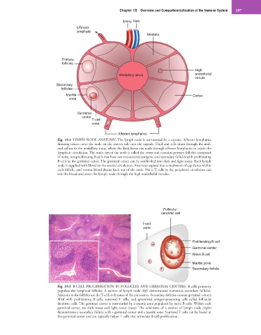
                            Fig. 19.4  LYMPH NODE ANATOMY. The lymph node is surrounded by a capsule. Afferent lymphatics
                            draining tissues enter the node on the convex side into the capsule. Fluid and cells drain through the node
                            and collect in the medullary sinus, where the fluid leaves the node through efferent lymphatics to rejoin the
                            lymphatic circulation. The outer rim of the node is called the cortex and contains primary follicles composed
                            of naive, nonproliferating B cells that have not encountered antigens, and secondary follicles with proliferating
                            B cells in the germinal center. The germinal center can be subdivided into dark and light zones. Each lymph
                            node is supplied with blood by the arterial circulation. Arterioles expand into a meshwork of capillaries within
                            each follicle, and venous blood drains back out of the node. Naive T cells in the peripheral circulation can
                            exit the blood and enter the lymph node through the high endothelial venules.


                                                                              Follicular
                                                                             dendritic cell

                                                                    T-cell
                                                                    zone


                                                                                           Proliferating B cell
                                                                                           Germinal center
                                                                                           Naive B cell
                                                                                           Mantle zone
                                                                                           Secondary follicle


                            Fig. 19.5  B-CELL PROLIFERATION IN FOLLICLES AND GERMINAL CENTERS. B cells primarily
                            populate the lymphoid follicles. A section of lymph node (left) demonstrates numerous secondary follicles.
                            Adjacent to the follicles are the T cell–rich zones of the paracortex. Secondary follicles contain germinal centers
                            filled  with  proliferating  B  cells,  scattered T  cells,  and  specialized  antigen-presenting  cells  called  follicular
                            dendritic cells. The germinal center is surrounded by a mantle zone populated by naive B cells. Within each
                            germinal  center,  are  dark  zones  and  light  zones  (inset). The  schematic  of  a  section  of  lymph  node  (right)
                            demonstrates a secondary follicle with a germinal center and a mantle zone. Scattered T cells can be found in
                            the germinal center and are typically helper T cells that stimulate B-cell proliferation.
   250   251   252   253   254   255   256   257   258   259   260