
Page 2627 - Hematology_ Basic Principles and Practice ( PDFDrive )
P. 2627
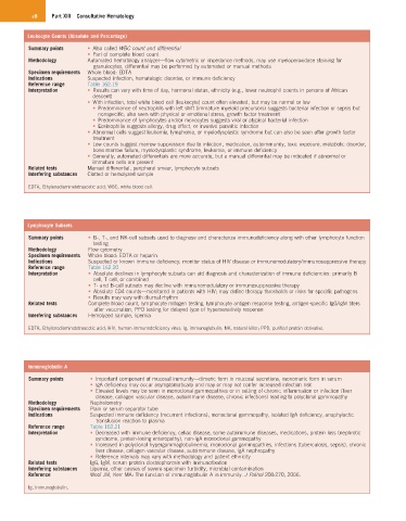
e8    Part XIII  Consultative Hematology


         Leukocyte Counts (Absolute and Percentage)
          Summary points   •  Also called WBC count and differential
                           •  Part of complete blood count
          Methodology      Automated hematology analyzer—flow cytometric or impedance methods, may use myeloperoxidase staining for
                             granulocytes, differential may be performed by automated or manual methods
          Specimen requirements  Whole blood: EDTA
          Indications      Suspected infection, hematologic disorder, or immune deficiency
          Reference range  Table 162.19
          Interpretation   •  Results can vary with time of day, hormonal status, ethnicity (e.g., lower neutrophil counts in persons of African
                             descent)
                           •  With infection, total white blood cell (leukocyte) count often elevated, but may be normal or low
                             •  Predominance of neutrophils with left shift (immature myeloid precursors) suggests bacterial infection or sepsis but
                               nonspecific; also seen with physical or emotional stress, growth factor treatment
                             •  Predominance of lymphocytes and/or monocytes suggests viral or atypical bacterial infection
                             •  Eosinophilia suggests allergy, drug effect, or invasive parasitic infection
                           •  Abnormal cells suggest leukemia, lymphoma, or myelodysplastic syndrome but can also be seen after growth factor
                             treatment
                           •  Low counts suggest marrow suppression due to infection, medication, autoimmunity, toxic exposure, metabolic disorder,
                             bone marrow failure, myelodysplastic syndrome, leukemia, or immune deficiency
                           •  Generally, automated differentials are more accurate, but a manual differential may be indicated if abnormal or
                             immature cells are present
          Related tests    Manual differential, peripheral smear, lymphocyte subsets
          Interfering substances  Clotted or hemolyzed sample
          EDTA, Ethylenediaminetetraacetic acid; WBC, white blood cell.


         Lymphocyte Subsets
          Summary points    •  B-, T-, and NK-cell subsets used to diagnose and characterize immunodeficiency along with other lymphocyte function
                             testing
          Methodology       Flow cytometry
          Specimen requirements  Whole blood: EDTA or heparin
          Indications       Suspected or known immune deficiency; monitor status of HIV disease or immunomodulatory/immunosuppressive therapy
          Reference range   Table 162.20
          Interpretation    •  Absolute declines in lymphocyte subsets can aid diagnosis and characterization of immune deficiencies: primarily B
                             cell, T cell, or combined
                            •  T- and B-cell subsets may decline with immunomodulatory or immunosuppressive therapy
                            •  Absolute CD4 counts—monitored in patients with HIV; may define therapy thresholds or risks for specific pathogens
                            •  Results may vary with diurnal rhythm
          Related tests     Complete blood count, lymphocyte mitogen testing, lymphocyte antigen response testing, antigen-specific IgG/IgM titers
                             after vaccination, PPD testing for delayed type of hypersensitivity response
          Interfering substances  Hemolyzed sample, lipemia
          EDTA, Ethylenediaminetetraacetic acid; HIV, human immunodeficiency virus; Ig, immunoglobulin; NK, natural killer; PPD, purified protein derivative.


         Immunoglobulin A
          Summary points    •  Important component of mucosal immunity—dimeric form in mucosal secretions, monomeric form in serum
                            •  IgA deficiency may occur asymptomatically and may or may not confer increased infection risk
                            •  Elevated levels may be seen in monoclonal gammopathies or in setting of chronic inflammation or infection (liver
                              disease, collagen vascular disease, autoimmune disease, chronic infections) leading to polyclonal gammopathy
          Methodology       Nephelometry
          Specimen requirements  Plain or serum separator tube
          Indications       Suspected immune deficiency (recurrent infections), monoclonal gammopathy, isolated IgA deficiency, anaphylactic
                              transfusion reaction to plasma
          Reference range   Table 162.21
          Interpretation    •  Decreased with immune deficiency, celiac disease, some autoimmune diseases, medications, protein loss (nephrotic
                              syndrome, protein-losing enteropathy), non-IgA monoclonal gammopathy
                            •  Increased in polyclonal hypergammaglobulinemia, monoclonal gammopathies, infections (tuberculosis, sepsis), chronic
                              liver disease, collagen vascular disease, autoimmune disease, IgA nephropathy
                            •  Reference intervals may vary with methodology and patient ethnicity
          Related tests     IgG, IgM, serum protein electrophoresis with immunofixation
          Interfering substances  Lipemia, other causes of severe specimen turbidity, microbial contamination
          Reference         Woof JM, Kerr MA: The function of immunoglobulin A in immunity. J Pathol 208:270, 2006.

          Ig, Immunoglobulin.