
Page 355 - Hematology_ Basic Principles and Practice ( PDFDrive )
P. 355
Chapter 25 Tolerance and Autoimmunity 291
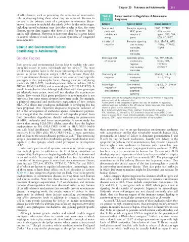
of self-tolerance, such as preventing the activation of autoreactive TABLE Genes Involved in Regulation of Autoimmune
cells or downregulating them when they are activated. Because in 25.2 Responses
no case is the primary cause of a polygenic autoimmune disease
known, it cannot be excluded that subtle defects in the earlier stages, Category Types of Genes a Known Examples b
including central tolerance, may also play a role; in fact, for some Central and Receptor signaling, CD45, PTPN22,
diseases, recent data suggest that there is a role for more “leaky” peripheral MHC genes, HLA (certain
central self-tolerance. However, it does seem clear that a gross defect deletion and receptor V types), CD3, CD4,
in central tolerance would lead to a severe syndrome of congenital anergy genes CD8, CD28/B7
autoimmunity.
Initiation of Receptor signaling, BLK, STAT-4, IRF5,
response costimulatory ITGAM, PTPN22,
Genetic and Environmental Factors molecules, FcγRII
Contributing to Autoimmunity adhesion
molecules
Genetic Factors Downregulation Apoptosis genes, Fas, TNF, CTLA4,
of response interleukins, CD40, CD3,
Both genetic and environmental factors help to explain why auto- negative TNFAIP3,
24
immunity occurs in some individuals and not others. The most costimulatory CD28/B7
well-known genetic factor is the major histocompatibility complex, molecules
known as human leukocyte antigen (HLA) in humans. Many dif- Channeling of Interleukins, STAT-4, IL-4, IL-10,
ferent autoimmune diseases are more or less associated with specific response interleukin IL-12, IFN-γ
genotypes at this polymorphic locus. Among these are ankylosing receptors
spondylitis (HLA-B27), insulin-dependent diabetes mellitus (HLA- Autoantigen Complement C1q, C2, C4, DNAse
DR3/4), RA (HLA-DR4), and to some degree SLE (HLA-DR2/3). It metabolism components, I, MER
should be emphasized that although individuals with these genotypes and apoptosis apoptosis
are relatively more prone, most will not develop the autoimmune signaling
disease. How certain HLA genes predispose to autoimmunity is not
very clear, but some recent advances have been made. For example, a Indicates some of the categories of genes that may be involved in regulating
a potential structural and mechanistic explanation of how certain autoimmunity at the indicated step.
Some genes in the categories of genes that may be involved in regulating
b
HLA-DR4 alleles may predispose individuals to developing RA has autoimmunity are indicated in the left column. Some have also been directly
been proposed. One important diagnostic/prognostic indicator of shown to play a role in autoimmunity.
RA is the development of antibodies against citrullinated peptides. BLK, B-lymphocyte kinase; CTLA, cytotoxic T-lymphocyte activation;
It is thought that the citrullination of self-peptides protect them IFN, interferon; IL, interleukin; IRF, interferon response factor; MER, C-mer
tyrosine kinase; MHC, major histocompatibility complex; PTP, posttransfusion
from proteolytic degradation, thereby enhancing its presentation purpura; STAT, signal transducers and activators of transcription.
of MHC molecules and hence autoreactivity. A recent study has
shown that among HLA-DR4 alleles, ones that have the highest
predisposition to RA (HLA-DRB1-04:01 or HLA-DRB1-04:04)
can only bind citrullinated Vimentin peptide, whereas the more these mutations lead to an age-dependent autoimmune syndrome
protective HLA-DR4 allele HLA-DRB1-04:02 is more permissive with autoantibody profiles that remarkably resemble human SLE,
25
and can bind to the uncitrullinated peptide as well. Thus restricting presumably, as a result of failure to eliminate postactivation T and
antigen presentation of this citrullinated peptide would lead to more B cells by the Fas-based mechanism. Exactly how defects in the
exposure to this epitope, which could predispose to development apoptotic Fas pathway lead to autoimmunity has yet to be elucidated.
of RA. Interestingly, a rare syndrome in humans with incomplete pen-
Inheritance patterns of all systemic autoimmune diseases suggest etrance, called autoimmune lymphoproliferation syndrome (ALPS),
that multiple genes, in addition to the HLA locus, contribute to has been traced to mutations in human Fas. Patients with ALPS
susceptibility. Such genes are beginning to be identified in human and develop polyclonal expansion of their lymphocytes and often display
in animal models. Interestingly, risk alleles have been identified for autoimmune cytopenias and less commonly SLE. The phenotypes of
a number of the same genes in more than one autoimmune disease; mutations in the Fas pathway illustrate two important points. They
these include CTLA-4, STAT-4, PTPN-22, TNFAIP3, and IRF-5, demonstrate the critical nature of the late downregulatory controls
all of which are known to regulate inflammation. Interestingly, most in preventing autoimmune disease. They also point out pathways
of them seem to have direct effects on B-cell function or activity. in which less severe mutations might be discovered that account for
Table 25.2 lists categories of genes that are likely involved in genetic human disease.
predisposition to autoimmune disease, drawing from both human A final category of genes regulates the clearance of self-antigens and
and murine studies. Note that these include genes involved in the dead cells, which is particularly important in systemic autoimmune
processes of antigen sequestration, T–B collaboration, and immune diseases such as SLE. These include complement components C4,
response downregulation that were discussed earlier as key features C3, and C2, C1q, and genes such as MER, which plays a role in
of the self-tolerance mechanisms that normally prevent autoimmune signaling for the uptake of apoptotic fragments by macrophages.
disease. In ongoing work, the precise nature of defects in these Evidently, when self-antigens are not cleared promptly after cell
genes may be defined; these include noncoding polymorphisms death, they can become targets of the immune system, leading to
that affect expression levels in addition to structural alleles. This autoimmunity to intracellular components such as chromatin.
will in turn permit screening for defects in human autoimmune As noted, TLRs can recognize some of these molecules when they
disease patients with the ultimate goals of aiding diagnosis, providing are present in high concentrations, thus providing proinflammatory
insights into pathogenic mechanisms, and guiding patient-specific signals. In murine models of lupus, it has been demonstrated in vivo
therapies. that TLR9 is required to generate antichromatin autoantibodies and
Although human genetic studies and animal models suggest that TLR7, which recognizes RNA, is required for the generation of
26
multigenic inheritance, there are certain instructive cases in which autoantibodies to RNA-related antigens. Indeed, a mutant mouse
single-gene defects play a major role. A well-studied example of muta- with a double dose of TLR7 develops spontaneous lupus with high
tions in these genes is the lpr/lpr mouse, which carries an inactivated levels of RNA antibodies. Stimulation of these TLRs on special-
17
murine Fas. The gld mutation, which inactivates murine Fas ligand ized plasmacytoid dendritic cells leads to release of abundant type
18
(FasL), has a very similar phenotype to the lpr/lpr mouse. Both of I interferon, which itself may be causally linked to lupus in mice