
Page 406 - Hematology_ Basic Principles and Practice ( PDFDrive )
P. 406
328 Part IV Disorders of Hematopoietic Cell Development
−/−
Abrogation of PU.1 expression in PU.1 mice results in peri- exhibit high DNA-binding affinity. It has further been suggested
natal lethality accompanied by the absence of mature monocytes/ that CDP-binding activity is restricted to proliferating cells, in
macrophages and B cells and delayed and reduced granulopoiesis. which CDP target genes are repressed. These targets are upregulated
Following in vitro differentiation, embryonic stem (ES) cells derived as cells undergo cell cycle arrest and terminal differentiation, in
−/−
from PU.1 blastocysts fail to express mature myeloid cell markers, association with a decrease in CDP binding. Target genes of CDP
suggesting that PU.1 is not essential for the initial events associated include c-myc, c-mos, and the thymidine kinase (TK), cdk inhibi-
with myeloid lineage commitment but is necessary for the later stages tor p21(WAF1/CIP1), cystic fibrosis transmembrane conductance
of development. regulator (CFTR), transforming growth factor-β (TGF-β) type II
receptor, gp91 phox, major histocompatibility complex (MHC) class
Growth Factor Independence-1 (Gfi-1) I locus, and neutrophil SGP genes.
The Gfi-1 gene was first identified as a target of proviral insertion During myeloid differentiation, CDP binding has been shown
following infection with Moloney murine leukemia virus (MoMuLV) to regulate genes that are expressed at widely disparate stages of
resulting in interleukin-2 (IL-2) factor independence in a rat lym- differentiation. For example, it represses the gp91 phox gene, which
20
phoma cell line (reviewed by van der Meer et al ). Gfi-1 is a highly is expressed at a much earlier time in myelopoiesis than is the case
conserved gene that encodes a 55-kDa nuclear proto-oncogene that for the LF gene. The mechanism by which CDP mediates repression,
harbors six C 2H 2 type zinc finger domains at the C-terminus and and the means by which it modulates stage-specific gene expres-
a 20–amino acid stretch at the N-terminus known as the SNAG sion at different stages of differentiation within a single lineage, are
20
domain (reviewed by van der Meer et al ). The SNAG domain not fully understood. CDP is reported to have repressive activity
that appears to be conserved in the Snail/Slug family of proteins, associated with its ability to be displaced by a positive trans-acting
has been shown to confer transcriptional repressor activity on Gfi-1. factor involving the CR1 and CR2 cut repeats. However, other
The human Gfi-1 gene is located on chromosome 1p22 and its modes of repressive activity involving the two active repression
closely related paralog Gfi1b maps to chromosome 9q34. Gfi-1 is domains within the C terminus of CDP have also been reported.
expressed at high levels in the thymus and BM while Gfi1B expres- CDP has been shown to function as a repressor of transcription via
sion is confined to the BM and spleen. Homozygous knockout of chromatin modification through recruitment of histone deacetylases
Gfi-1B results in embryonic lethality at day E15, despite the fact that (HDACs), consistent with the hypothesis that transcriptional silenc-
myelopoiesis is normal. Death in these mice has been attributed to a ing is associated with hypoacetylated histones. Both acetylation and
failure of erythropoiesis and megakaryopoiesis. phosphorylation of CDP are posttranscriptional modifications that
The essential role of Gfi-1 in neutrophil differentiation became have been postulated to regulate CDP function. Thus differential
apparent following two reports of gene disruption in mice. Gfi-1-null modification, by phosphorylation or acetylation, of CDP-DNA
mice are severely neutropenic and eventually succumb to bacterial complexes binding the promoters of target genes could result in the
infections. In addition, these mice lack mature neutrophils and observed differential repression exerted by CDP during neutrophil
their granulocyte precursors are unable to differentiate into mature development.
neutrophils upon induction with G-CSF. These cells also lack SGP
−/−
−/−
expression reminiscent of C/EBP granulocytes. Gfi-1 BM con-
+
+
tains an atypical Gr1 Mac1 myeloid precursor cell that appeared to ROLE OF DEVELOPMENTALLY IMPORTANT
share characteristics of both granulocyte and macrophage precursors. NEUTROPHIL-SPECIFIC GENES IN DISEASE
−/−
Ectopic expression of Gfi-1 in ex vivo sorted Gfi1 progenitor cells
restores G-CSF–mediated neutrophil maturation to these cells. These Our understanding of the role of neutrophil-specific genes has been
observations provide evidence for the critical role of Gfi-1 in the enhanced by the study of mice in which targeted disruption of
neutrophil maturation program. Other studies have further demon- a gene results in phenotypically important defects in neutrophil
strated that Gfi-1 together with C/EBPε synergize to transactivate differentiation and function. Similarly, the importance of these
the promoters of late myeloid genes. This synergy is lost in a patient genes has been underscored by the analysis of naturally occurring
with SGD, who has a heterozygous substitution mutation in the C/ genetic events within these genes that result in human disease.
EBPε gene and decreased levels of Gfi-1 in the BM. The links between some genes and the diseases induced by their
Heterozygous dominant negative mutations in the Gfi-1 gene dysfunction may be anticipated by their important roles in neu-
have been described in two patients with SCN, underscoring the trophil differentiation and function, whereas the pathophysiologic
role of Gfi-1 in the neutrophil maturation pathway. It has been link between others and the diseases they induce remain elusive
suggested that mutant Gfi-1 in these patients alters the expression (Table 27.2).
of ELANE, mutations in which are commonly associated with SCN
(see later). This observation confirms the vital role Gfi-1 plays in
human granulopoiesis.
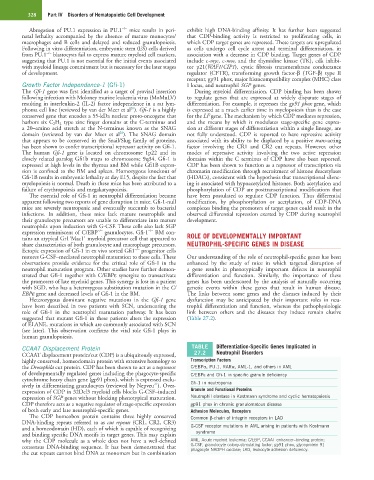
CCAAT Displacement Protein TABLE Differentiation-Specific Genes Implicated in
CCAAT displacement protein/cut (CDP) is a ubiquitously expressed, 27.2 Neutrophil Disorders
highly conserved, homeodomain protein with extensive homology to Transcription Factors
the Drosophila cut protein. CDP has been shown to act as a repressor C/EBPα, PU.1, RARα, AML-1, and others in AML
of developmentally regulated genes including the phagocyte-specific C/EBPε and Gfi-1 in specific granule deficiency
cytochrome heavy chain gene (gp91 phox), which is expressed exclu-
21
sively in differentiating granulocytes (reviewed by Nepveu ). Over- Gfi-1 in neutropenia
expression of CDP in 32Dcl3 myeloid cells blocks G-CSF–induced Granule and Functional Proteins
expression of SGP genes without blocking phenotypical maturation. Neutrophil elastase in Kostmann syndrome and cyclic hematopoiesis
CDP therefore acts as a negative regulator of stage-specific expression gp91 phox in chronic granulomatous disease
of both early and late neutrophil-specific genes. Adhesion Molecules, Receptors
The CDP homeobox protein contains three highly conserved Common β-chain of integrin receptors in LAD
DNA-binding repeats referred to as cut repeats (CR1, CR2, CR3)
and a homeodomain (HD), each of which is capable of recognizing G-CSF receptor mutations in AML arising in patients with Kostmann
and binding specific DNA motifs in target genes. This may explain syndrome
why the CDP molecule as a whole does not have a well-defined AML, Acute myeloid leukemia; C/EBP, CCAAT enhancer–binding protein;
consensus DNA-binding sequence. It has been demonstrated that G-CSF, granulocyte colony-stimulating factor; gp91 phox, glycoprotein 91
phagocyte NADPH oxidase; LAD, leukocyte adhesion deficiency.
the cut repeats cannot bind DNA as monomers but in combination