
Page 477 - Hematology_ Basic Principles and Practice ( PDFDrive )
P. 477
398 Part IV Disorders of Hematopoietic Cell Development
Laboratory support for the immune hypothesis first came from Acute Chronic
coculture experiments in which mononuclear cells from AA patients’
blood or BM were shown to suppress in vitro colony formation by 10 BMT conditioning
hematopoietic progenitor cells. T-cell depletion sometimes improved 100% mortality with routine medical support
colony formation in vitro. Patients’ blood and BM cells were shown
to produce a soluble factor that inhibited hematopoiesis, ultimately LD (human experience)
identified as interferon (IFN)-γ. Patients’ T cells overproduce IFN-γ 50
and tumor necrosis factor (TNF), two cytokines that inhibit hema- Radiation sickness
topoietic proliferation. Tbet, a transcriptional regulator that is critical Bone marrow hypoplasia
to Th1 polarization, is constitutionally expressed in a majority of AA LD (animal models)
50
patients. AA blood and BM also contains elevated numbers of acti- 1
vated cytotoxic lymphocytes, and activity and levels of these cytotoxic Leukemogenesis in
cells are decreased with antithymocyte globulin (ATG) therapy. T A-bomb survivors Increased
regulatory cells, as in other human immune-mediated diseases, are spontaneous
decreased in AA. IFN-γ and TNF negative effects on the proliferation mutation rate
of early and late hematopoietic progenitor and stem cells is far more
potent when these cytokines are secreted into the BM microenviron-
ment than when they were simply added to the cultures. IFN-γ and
TNF can suppress hematopoiesis by inhibiting cell proliferation, .1 Loss of glycophorin phenotype
inducing Fas-mediated apoptosis, and blocking hematopoietic growth
factor intracellular signals. The early immune system events that must
precede the global destruction of hematopoietic cells are not clear.
Involvement of CD4 lymphocytes has been suggested based on the
overrepresentation of HLA-DR15 among patients with immune-
mediated AA. Clones of HLA-DR–restricted T cells derived from a Dose, Gy
few patients have been shown to proliferate in response to BM cells.
Many features of human AA can be reproduced in mouse models .01
of GVHD in which the donor inoculum lacks stem cells. Major and
minor histocompatibility mismatch demonstrates the potency and Thyroid scan
specificity of small numbers of T cells, the role of cytokines, efficacy Maximal permissible
of immunosuppressive therapies, an “innocent bystander effect,” and occupational exposure/year
roles for specific lymphocyte regulatory and effector T cell subsets. 7 Liver-spleen scan
Radiation .001 CT scan 5000 feet natural
sea level radiation/year
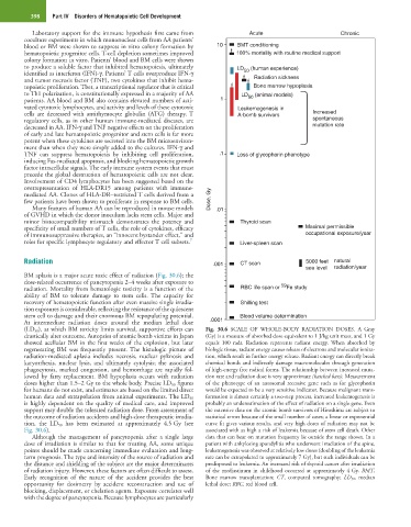
BM aplasia is a major acute toxic effect of radiation (Fig. 30.6); the
dose-related occurrence of pancytopenia 2–4 weeks after exposure to 59
radiation. Mortality from hematologic toxicity is a function of the RBC life span or Fe study
ability of BM to tolerate damage to stem cells. The capacity for
recovery of hematopoietic function after even massive single irradia- Shilling test
tion exposures is considerable, reflecting the resistance of the quiescent
stem cell to damage and their enormous BM repopulating potential. Blood volume determination
At intermediate radiation doses around the median lethal dose .0001
(LD 50 ), at which BM toxicity limits survival, supportive efforts can Fig. 30.6 SCALE OF WHOLE-BODY RADIATION DOSES. A Gray
drastically alter outcome. Autopsies of atomic bomb victims in Japan (Gy) is a measure of absorbed dose equivalent to 1 J/kg unit mass, and 1 Gy
showed acellular BM in the first weeks of the explosion, but later equals 100 rads. Radiation represents radiant energy. When absorbed by
regenerating BM was frequently present. The histologic picture of biologic tissue, radiant energy causes release of electrons and molecular ioniza-
radiation-mediated aplasia includes necrosis, nuclear pyknosis and tion, which result in further energy release. Radiant energy can directly break
karyorrhexis, nuclear lysis, and ultimately cytolysis; the associated chemical bonds and indirectly damage macromolecules through generation
phagocytosis, marked congestion, and hemorrhage are rapidly fol- of high-energy free radical forms. The relationship between increased muta-
lowed by fatty replacement. BM hypoplasia occurs with radiation tion rate and radiation dose is very approximate (hatched bars). Measurement
doses higher than 1.5–2 Gy to the whole body. Precise LD 50 figures of the phenotype of an autosomal recessive gene such as for glycophorin
for humans do not exist, and estimates are based on the limited direct would be expected to be a very sensitive indicator. Because malignant trans-
human data and extrapolation from animal experiments. The LD 50 formation is almost certainly a two-step process, increased leukemogenesis is
is highly dependent on the quality of medical care, and improved probably an underestimation of the effect of radiation on a single gene. Even
support may double the tolerated radiation dose. From assessment of the extensive data on the atomic bomb survivors of Hiroshima are subject to
the outcome of radiation accidents and high-dose therapeutic irradia- statistical errors because of the small number of cases; a linear or exponential
tion, the LD 50 has been estimated at approximately 4.5 Gy (see curve fit gives various results, and very high doses of radiation may not be
Fig. 30.6). associated with as high a risk of leukemia because of stem cell death. Other
Although the management of pancytopenia after a single large data that can bear on mutation frequency lie outside the range shown. In a
dose of irradiation is similar to that for treating AA, some unique patient with ankylosing spondylitis who underwent irradiation of the spine,
points should be made concerning immediate evaluation and long- leukemogenesis was observed at relatively low doses (doubling of the leukemia
term prognosis. The type and intensity of the source of radiation and rate can be extrapolated to approximately 7 Gy), but such individuals can be
the distance and shielding of the subject are the major determinants predisposed to leukemia. An increased risk of thyroid cancer after irradiation
of radiation injury. However, these factors are often difficult to assess. of the mediastinum in childhood occurred at approximately 4 Gy. BMT,
Early recognition of the nature of the accident provides the best Bone marrow transplantation; CT, computed tomography; LD 50, median
opportunity for dosimetry by accident reconstruction and use of lethal dose; RBC, red blood cell.
blocking, displacement, or chelation agents. Exposure correlates well
with the degree of pancytopenia. Because lymphocytes are particularly