
Page 484 - Hematology_ Basic Principles and Practice ( PDFDrive )
P. 484
Chapter 30 Aplastic Anemia 405
A B C DD E
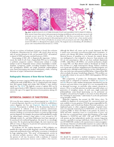
Fig. 30.9 MORPHOLOGY OF OTHER DISEASES THAT MAY MANIFEST WITH PANCYTOPENIA.
Bone marrow biopsy from patient with pancytopenia showing myelofibrosis and osteosclerosis associated with
metastatic prostate cancer (A). The aspirate was hypocellular but did show occasional tumor clusters (B).
Another case where the patient presented with pancytopenia and was found to have a bone marrow packed
with lymphoma cells (C). Hairy cell leukemia can present with pancytopenia and with a hypocellular bone
marrow (D) difficult to distinguish from aplastic anemia. The diagnosis rests on identifying a B-cell infiltrative
process with immunohistochemical stains (E, CD20).
AA and are evidence of aleukemic leukemia or herald the evolution although the blood cell counts can be severely depressed, the BM
+
of leukemia. Histochemistry for CD34 cells should show staining is much more commonly normo/hypercellular than hypoplastic. In
+
only of vascular elements in AA, and increased CD34 cell numbers patients with chronic BM failure, serial BM specimens may not be
is typical of MDS and acute myeloid leukemia (AML). identical because of sampling error or because the original disease
Karyotyping of BM cells is diagnostically important. Unfortu- was misdiagnosed or has changed its character. Some patients with
nately, the yield of cells from a hypocellular BM can be inadequate AA are not pancytopenic; they do not have uniform depressions
to perform cytogenetic analysis. Chromosome analysis is usually of RBC, white blood cell (WBC), and platelet production, despite
normal in AA but frequently reveals a clonal abnormality in myelo- an empty BM, and their clinical course is dominated by failure in
dysplasia. Cytogenetic studies, including interphase fluorescent in two cell lines or a single hematopoietic lineage. Related conditions
situ hybridization (FISH) and single nucleotide polymorphisms such as pure red cell aplasia, amegakaryocytic thrombocytopenia, and
array–based karyotyping may produce informative results, including agranulocytosis, although usually distinctive in their clinical presenta-
detection of cryptic chromosomal abnormalities. 10 tion, can evolve into more generalized BM failure. A hypocellular BM
often precludes the proper morphologic diagnosis. This problem can
be especially evident in the case of a MDS with hypoplastic BM (see
Radiographic Measures of Bone Marrow Function Table 30.7).
BM cytogenetics, if positive for chromosome abnormalities,
Magnetic resonance imaging (MRI) with spin-echo sequences can be usually leads to a diagnosis of leukemia or MDS (see Chapters 59
useful in the study of BM disease. On T1-weighted spin-echo images, and 60). However, some random chromosomal abnormalities may be
fatty BM appears bright and cellular BM exhibits a lower density transient and some believe that typical AA is not incompatible with
signal (see Fig. 30.9). The high-fat content of aplastic BM can be an abnormal karyotype, in particular when somatic mosaicism is
readily appreciated on MRI. Magnetic resonance spectroscopy, which present. Often, an acellular specimen precludes successful culture and
detects the type of fat signal, has shown diverse patterns among AA generation of metaphase smears. In such cases, single nucleotide
patients. polymorphisms arrays–based karyotyping can be performed on
interphase cells and may be helpful in detection of clonal abnormali-
ties. Screening for monosomy 7 and trisomy 8 can be also performed
DIFFERENTIAL DIAGNOSIS OF PANCYTOPENIA using interphase FISH.
Molecular diagnostics has entered the BM failure clinic. To
AA is not the most common cause of pancytopenia (see Table 30.1). establish the diagnosis of constitutional AA, commercial panels are
A rational diagnostic algorithm can be very helpful in establishing a now available to screen germline DNA for genes responsible for FA,
correct diagnosis (see box on Diagnostic Algorithm in Aplastic telomeropathy, and Schwachman-Bodian-Diamond syndrome, and
Anemia). Pancytopenia is unlikely to be the presenting feature of implicated genes in other congenital syndromes. Acquired muta-
hypersplenism in cirrhosis or of Evans syndrome (autoimmune tions of recurrently mutated genes in MDS/AML can be detected in
hemolytic anemia and thrombocytopenia) in systemic lupus erythe- circulating WBC in about one-third of AA patients. In contrast to
matosus. Findings on physical examination can point strongly toward MDS and AML, mutations occur in a limited subset of genes, and
another diagnosis. For example, the patient with myelofibrosis usually the clone size, as estimated from variant allele frequency, is also small.
has splenomegaly, whereas a large spleen is very unusual in AA. Mutations in DNMT3A and ASXL, CBL and SETBP1 correlate
Although vitamin B 12 and folate deficiencies have been reported to with patient age but independently predict for a poorer long-term
be associated with erythroid hypoplasia, this must be an exceedingly outcome, especially in younger individuals. Only mutations in PIGA
rare event. For the practicing hematologist, the most important and and BCOR/BCORL correlate with responsiveness to immunosuppres-
difficult choice of diagnoses in patients with pancytopenia is among sive therapy. Detection of unfavorable mutations in a patient who has
the primary BM disorders. failed treatment may influence the decision to undertake high risk or
In moderate AA, the modest depression of BM cellularity can alternative stem cell transplantation. 11
muddle the single most reliable diagnostic criterion. BM cellularity is
imprecisely quantitated at best, and further uncertainty is introduced
by large sampling errors. “Hot spots” of hematopoietic activity in TREATMENT
an otherwise acellular specimen reflect biologic heterogeneity in
the pattern of cell loss. In patients with a syndrome of transient AA should be considered a medical emergency. Lives are lost,
pancytopenia, spontaneous recovery occurs within a few months; mainly because the grave consequences of severe pancytopenia go