
Page 508 - Hematology_ Basic Principles and Practice ( PDFDrive )
P. 508
428 Part IV Disorders of Hematopoietic Cell Development
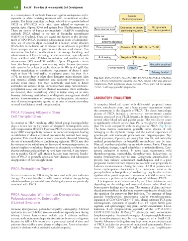
as a consequence of antibody formation against endogenous eryth- Anemia
ropoietin or while receiving treatment with recombinant erythro- Reticulocytopenia
poietin. The latter condition has been referred to as epoetin-induced
PRCA or EPO-PRCA with initial cases related to exposure to
epoetin alpha (Eprex; 92%) and epoetin beta (NeoRecormon; 8%). Decreased or absent No dysplasia
There are reports of human erythropoietin (HuEPO) neutralizing Bone marrow exam erythroid precursors Normal cytogenetics
antibody PRCA related to the use of biosimilar recombinant
HuEPO in Thailand. There are several risk factors to the develop- Rule out
ment of EPO-PRCA, including subcutaneous route of administra- drugs
tion, use of epoetin alpha stabilized in a human serum albumin systemic diseases
(HSA)–free formulation, use of silicone oil as lubricant in prefilled Flow cytometry Absence of CLL, NK-LGL, or T-LGL
Eprex syringes, and use in patients with chronic renal disease. This
observation has led to modification in the storage, handling, and
administration of Eprex favoring IV administration, especially with
Eprex stabilized with HSA-free formulation, and avoidance of the B19 testing Negative PCR, serology
subcutaneous (SC) non–HSA stabilized Eprex. Diagnostic criteria
have also been proposed incorporating major features (treatment Idiopathic acquired
with epoetin for at least 3 weeks, decrease in Hb by 0.1 g/dL/day Family history PRCA
without transfusions or transfusion requirement of about 1 unit/
week to keep Hb level stable, reticulocyte count less than 10 × Primary familial
9
10 /L, no major drop in other blood lineages), minor features (skin Fig. 32.3 DIAGNOSTIC ALGORITHM IN PURE RED CELL APLASIA.
and systemic allergic reactions), and accessory investigations to CLL, Chronic lymphocytic leukemia; NK-LGL, natural killer large granular
exclude other causes. The most commonly used tests to detect lymphocyte; PCR, polymerase chain reaction; PRCA, pure red cell aplasia;
antibodies are enzyme-linked immunosorbent assay, radioimmuno- T-LGL, T-cell large granular lymphocyte.
precipitation assay, and surface plasmon resonance. Once antibodies
are detected, their neutralizing ability is tested using an in vitro
bioassay. Following establishment of diagnosis, management should
include discontinuation of exogenous erythropoietin, administra- LABORATORY EVALUATION
tion of immunosuppressive agents, or, in cases of anemia secondary
to renal insufficiency, renal transplantation. A complete blood cell count with differential, peripheral smear
review, reticulocyte count, and a bone marrow examination remain
PRCA Following Allogeneic Stem the cornerstone in the diagnosis of PRCA. The classic hematologic
picture of PRCA includes a normocytic, normochromic anemia
Cell Transplantation (anemia associated with T-LGL leukemia is often macrocytic) with a
normal white blood cell and platelet count. The reticulocyte count
In contrast to HLA matching, ABO blood group incompatibility is significantly reduced to less than 1% (a reticulocyte level greater
plays a minor role in the success of allogeneic hematopoietic stem than 2% is not compatible with the diagnosis of PRCA (Fig. 32.3).
cell transplantation (HSCT). However, PRCA may be associated with The bone marrow examination generally shows absence of cells
major ABO incompatibility between the donor and recipient, leading belonging to the erythroid lineage and the normal appearance of
to inhibition of donor erythroid precursors by residual host isoag- granulocytic and monocytic precursors and megakaryocytes. Ery-
glutinins. This complication is more commonly observed following throid precursors, if present, are usually less than 1%, and only a few
the use of nonmyeloablative conditioning regimens. PRCA may also residual proerythroblasts or basophilic erythroblasts may be seen.
be resistant to the withdrawal or decrease of immunosuppression, or Blast cell numbers and cellularity are within normal limits. There are
donor lymphocyte infusions. Responses to rituximab, erythropoietin, no dysplastic changes, ringed sideroblasts, or reticulin fibrosis. Cyto-
plasma exchange and azathioprine have been reported. A case respon- genetic evaluation is normal. In some cases, neutropenia, mild
+
sive to purified CD34 cell infusion has also been reported. Resolu- thrombocytopenia, eosinophilia, thrombocytosis, leukocytosis, or
tion of PRCA is generally associated with decrease and subsequent relative lymphocytosis may be seen. Cytogenetic abnormalities, if
disappearance of host isoagglutinins. present, may indicate concomitant myelodysplasia and is a poor
prognostic marker for both response to treatment and propensity to
leukemic transformation. During the course of PRCA patients, inef-
PRCA Postradiation Therapy fective erythropoiesis characterized by a maturation arrest at the
proerythroblast or basophilic erythroblast stage may be observed and
In rare circumstances, PRCA may be associated with prior radiation signifies either partial response to treatment or initial recovery from
therapy. The cases described usually involve radiation therapy being treatment or a prelude to the development of full-blown PRCA.
administered to a patient with an underlying thymoma not previously It is important to exclude vitamin B 12 and folate deficiencies, and
associated with PRCA. depending on the etiology and associated disease, other blood and
bone marrow findings may be seen. The presence of giant and vacu-
olated pronormoblasts in the bone marrow examination should raise
PRCA Associated With Immune Dysregulation, the suspicion for parvovirus B19 infection. The presence of large
Polyendocrinopathy, Enteropathy, granular lymphocytosis, neutropenia, and/or thrombocytopenia,
+
+
+
expansion of CD3 CD8 CD57 T cells, clonal cytotoxic TCR gene
X-Linked Syndrome rearrangement, expansion of specific TCR Vβ region family gene
segment, and splenomegaly may point toward concomitant T-LGL
+
+
Immune dysregulation, polyendocrinopathy, enteropathy, X-linked leukemia, B-cell lymphocytosis, especially of the CD5 /CD19 /
+
+
−
syndrome is a rare X-linked recessive condition typically seen during CD20 /CD23 /cyclin D /SmIg −dim phenotype with concomitant
infancy. Clinical features may include type 1 diabetes mellitus, lymphadenopathy, hepatosplenomegaly, hypogammaglobulinemia,
eczema, and autoimmune hepatitis. Anemia can be severe at diagnosis and thrombocytopenia may be very suggestive of a B-cell CLL.
because the fall in Hb occurs over a protracted period of time and Another laboratory finding that may help point to a secondary cause
patients often exhibit a good degree of adaptation. Arrest of erythro- of PRCA includes the presence of monoclonal gammopathy. Parvo-
poiesis is obvious with a profound reticulocytopenia. virus B19 DNA titers (DNA hybridization and amplification