
Page 565 - Hematology_ Basic Principles and Practice ( PDFDrive )
P. 565
480 Part V Red Blood Cells
A B C D E
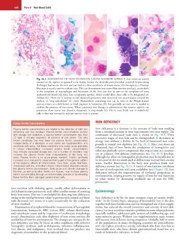
Fig. 36.2 ASSESSMENT OF IRON STORES ON A BONE MARROW ASPIRATE. Iron stores are usually
assessed on the aspirate as opposed to the biopsy because the decalcification procedure required for processing
the biopsy leaches out the iron and can lead to a false conclusion of absent stores. On the aspirate, a Prussian
blue stain is usually used to evaluate iron. This can demonstrate iron stores (blue reaction product), particularly
in the cytoplasm of macrophages and histiocytes (A–B). Iron can also be seen in the cytoplasm of some
nucleated red blood cells (tiny blue cytoplasmic specks), which would allow these cells to be designated sid-
eroblasts (C). These are in contrast to red blood cell precursors with abnormal iron accumulation around the
nucleus, or “ring sideroblasts” (C, inset). Hemosiderin containing iron can be seen on the Wright-stained
aspirate smears as a dark brown or black pigment in histiocytes (D), but generally an iron stain is needed to
confirm the presence of iron stores. When parenteral iron therapy is administered, the marrow aspirate can
sometimes show coarse iron deposits, frequently in long streaks (E). This is most likely iron in endothelial
cells; it does not necessarily indicate marrow iron is present.
IRON DEFICIENCY
Plasma Ferritin Concentrations
Plasma ferritin concentrations are helpful in the detection of both iron Iron deficiency is a decrease in the amount of body iron resulting
deficiency and iron overload. Plasma ferritin concentrations decline from a sustained increase in iron requirements over iron supply. The
with storage iron depletion; a plasma ferritin concentration less than continuum of decreased body iron is shown in Fig. 36.1. Three
12 mg/L is virtually diagnostic of absence of iron stores. The only successive stages of iron lack can be distinguished. A decrement in
known conditions that may lower the plasma ferritin concentration storage iron without a decline in the level of functional iron com-
independently of a decrease in iron stores are hypothyroidism and pounds is termed iron depletion (see Fig. 36.1). After iron stores are
ascorbate deficiency, but these conditions only rarely cause problems exhausted, lack of iron limits the production of hemoglobin and
in clinical interpretation. Increased plasma ferritin concentrations
may indicate increased storage iron, but a number of disorders may other metabolically active compounds that require iron as a constitu-
increase the plasma ferritin level independently of the body iron ent or cofactor. Iron-deficient erythropoiesis (see Fig. 36.1) develops,
store. Plasma ferritin is an acute-phase reactant. Ferritin synthesis although the effect on hemoglobin production may be insufficient to
increases as a nonspecific response that is part of the general pattern be detected by the standards used to differentiate normal from anemic
of the systemic effects of inflammation. Thus fever, acute infections, states. Further diminution in the body iron produces frank iron-
rheumatoid arthritis, and other chronic inflammatory disorders elevate deficiency anemia (see Fig. 36.1). A variety of mechanisms coordinate
the plasma ferritin concentration. Both acute and chronic damage to the rate of erythropoiesis with iron availability (see Chapter 35). Iron
the liver, as well as to other ferritin-rich tissues, may increase plasma deficiency reduces the responsiveness of erythroid progenitors to
ferritin concentration through an inflammatory process or by releasing erythropoietin, helping preserve the supply of iron for vital functions
tissue ferritins from damaged parenchymal cells.
in other tissues by decreasing erythroid use of iron for RBC
production.
iron excretion with chelating agents, usually either deferoxamine or
diethylenetriamine pentaacetic acid, offers another means of assessing Epidemiology
body iron stores. This test is not helpful for detecting iron deficiency,
owing to the overlap between values in persons with normal and those Iron deficiency is by far the most common cause of anemia world-
5
with decreased iron stores; it is used occasionally for the evaluation wide. In the United States, adequacy of bioavailable iron in the diet,
of iron overload. together with food fortification and the widespread use of iron supple-
Examination of peripheral blood by measurements of hemoglobin ments, has reduced the overall prevalence and severity of iron defi-
concentration, hematocrit, RBC indices, RBC volume distribution, ciency, but iron nutrition remains a problem in some subpopulations,
and reticulocyte count and by inspection of erythrocyte morphology especially toddlers, adolescent girls, women of childbearing age, and
reveals abnormalities only after depletion of iron stores restricts the some minority groups. Without iron supplementation, most women
availability of iron for erythropoiesis. The changes are not specific for will become iron-deficient during pregnancy. Globally, half or more
iron deficiency and may be found in other conditions with defective of the populations in many developing countries are iron-deficient,
hemoglobin synthesis, such as thalassemia, infection, inflammation, with the highest prevalence among individuals who have diets low in
liver disease, and malignancy. Iron overload does not produce any bioavailable iron, who have chronic gastrointestinal blood loss as a
diagnostic abnormalities in the peripheral blood. result of helminthic infection, or both. 5