
Page 584 - Hematology_ Basic Principles and Practice ( PDFDrive )
P. 584
Chapter 38 Heme Biosynthesis and Its Disorders 499
pre-ALAS1 pre-ALAS2
ALA ALA
Translation Translation
ALAS1 ALAS2
- -?
Glycine + Glycine +
pre−ALAS1 succinyl CoA pre−ALAS2 succinyl CoA
mRNA mRNA
Heme + Heme
+
Fe 2+ - Fe 2+
Protoporphyrin IX Protoporphyrin IX
Degradation
mfrn mfrn
Iron Iron
A pool B pool
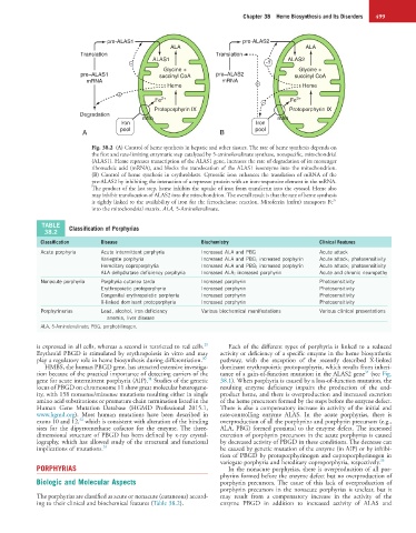
Fig. 38.2 (A) Control of heme synthesis in hepatic and other tissues. The rate of heme synthesis depends on
the first and rate-limiting enzymatic step catalyzed by 5-aminolevulinate synthase, nonspecific, mitochondrial
(ALAS1). Heme represses transcription of the ALAS1 gene, increases the rate of degradation of its messenger
ribonucleic acid (mRNA), and blocks the translocation of the ALAS1 isoenzyme into the mitochondrion.
(B) Control of heme synthesis in erythroblasts. Cytosolic iron enhances the translation of mRNA of the
pre-ALAS2 by inhibiting the interaction of a repressor protein with an iron-responsive element in the mRNA.
The product of the last step, heme inhibits the uptake of iron from transferrin into the cytosol. Heme also
may inhibit translocation of ALAS2 into the mitochondrion. The overall result is that the rate of heme synthesis
2+
is tightly linked to the availability of iron for the ferrochelatase reaction. Mitoferrin (mfrn) transports Fe
into the mitochondrial matrix. ALA, 5-Aminolevulinate.
TABLE Classification of Porphyrias
38.2
Classification Disease Biochemistry Clinical Features
Acute porphyria Acute intermittent porphyria Increased ALA and PBG Acute attack
Variegate porphyria Increased ALA and PBG; increased porphyrin Acute attack; photosensitivity
Hereditary coproporphyria Increased ALA and PBG; increased porphyrin Acute attack; photosensitivity
ALA dehydratase deficiency porphyria Increased ALA; increased porphyrin Acute and chronic neuropathy
Nonacute porphyria Porphyria cutanea tarda Increased porphyrin Photosensitivity
Erythropoietic protoporphyria Increased porphyrin Photosensitivity
Congenital erythropoietic porphyria Increased porphyrin Photosensitivity
X-linked dominant protoporphyria Increased porphyrin Photosensitivity
Porphyrinurias Lead, alcohol, iron deficiency Various biochemical manifestations Various clinical presentations
anemia, liver disease
ALA, 5-Aminolevulinate; PBG, porphobilinogen.
29
is expressed in all cells, whereas a second is restricted to red cells. Each of the different types of porphyria is linked to a reduced
Erythroid PBGD is stimulated by erythropoiesis in vitro and may activity or deficiency of a specific enzyme in the heme biosynthetic
play a regulatory role in heme biosynthesis during differentiation. 30 pathway, with the exception of the recently described X-linked
HMBS, the human PBGD gene, has attracted extensive investiga- dominant erythropoietic protoporphyria, which results from inheri-
34
tion because of the practical importance of detecting carriers of the tance of a gain-of-function mutation in the ALAS2 gene (see Fig.
31
gene for acute intermittent porphyria (AIP). Studies of the genetic 38.1). When porphyria is caused by a loss-of-function mutation, the
locus of PBGD on chromosome 11 show great molecular heterogene- resulting enzyme deficiency impairs the production of the end-
ity, with 158 nonsense/missense mutations resulting either in single product heme, and there is overproduction and increased excretion
amino acid substitutions or premature chain termination listed in the of the heme precursors formed by the steps before the enzyme defect.
Human Gene Mutation Database (HGMD Professional 2015.1, There is also a compensatory increase in activity of the initial and
www.hgmd.org). Most human mutations have been described in rate-controlling enzyme ALAS. In the acute porphyrias, there is
32
exons 10 and 12, which is consistent with alteration of the binding overproduction of all the porphyrins and porphyrin precursors (e.g.,
sites for the dipyrromethane cofactor for the enzyme. The three- ALA, PBG) formed proximal to the enzyme defect. The increased
dimensional structure of PBGD has been defined by x-ray crystal- excretion of porphyrin precursors in the acute porphyrias is caused
lography, which has allowed study of the structural and functional by decreased activity of PBGD in these conditions. The decrease can
implications of mutations. 33 be caused by genetic mutation of the enzyme (in AIP) or by inhibi-
tion of PBGD by protoporphyrinogen and coproporphyrinogen in
variegate porphyria and hereditary coproporphyria, respectively. 35
PORPHYRIAS In the nonacute porphyrias, there is overproduction of all por-
phyrins formed before the enzyme defect but no overproduction of
Biologic and Molecular Aspects porphyrin precursors. The cause of this lack of overproduction of
porphyrin precursors in the nonacute porphyrias is unclear, but it
The porphyrias are classified as acute or nonacute (cutaneous) accord- may result from a compensatory increase in the activity of the
ing to their clinical and biochemical features (Table 38.2). enzyme PBGD in addition to increased activity of ALAS and