
Page 585 - Hematology_ Basic Principles and Practice ( PDFDrive )
P. 585
500 Part V Red Blood Cells
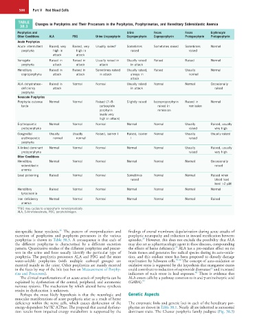
TABLE Changes in Porphyrins and Their Precursors in the Porphyrias, Porphyrinurias, and Hereditary Sideroblastic Anemia
38.3
Porphyrias and Urine Feces Feces Erythrocyte
Other Conditions ALA PBG Urine Uroporphyrin Coproporphyrin Coproporphyrin Protoporphyrin Protoporphyrin
Acute Porphyrias
Acute intermittent Raised, very Raised, very Usually raised a Sometimes Sometimes raised Sometimes Normal
porphyria high in high in raised raised
attack attack
Variegate Raised in Raised in Usually raised in Usually raised Raised Raised Normal
porphyria attack attack attack in attack
Hereditary Raised in Raised in Sometimes raised Usually raised, Raised Usually Normal
coproporphyria attack attack in attack always in normal
attack
ALA dehydratase– Raised in Normal Normal Usually raised Normal Normal Occasionally
deficiency attack in attack raised
porphyria
Nonacute Porphyrias
Porphyria cutanea Normal Normal Raised (7-/8- Slightly raised Isocoproporphyrin Raised in Normal
tarda carboxylate raised in remission
porphyrin remission
levels very
high in attack)
Erythropoietic Normal Normal Normal Normal Normal Usually Raised, usually
protoporphyria raised very high
Congenital Usually Usually Raised, isomer I Raised, isomer Normal Usually Usually raised
erythropoietic normal normal I raised
porphyria
X-linked dominant Normal Normal Normal Normal Normal Usually Raised, usually
protoporphyria raised very high
Other Conditions
Hereditary Normal Normal Normal Normal Normal Normal Occasionally
sideroblastic raised
anemia
Lead poisoning Raised Normal Normal Sometimes Normal Normal Raised when
raised blood lead
level >2 µM
Hereditary Raised Normal Normal Normal Normal Normal Normal
tyrosinemia
Iron deficiency Normal Normal Normal Normal Normal Normal Raised
anemia
a PBG may cyclize to uroporphyrin nonenzymatically.
ALA, 5-Aminolevulinate; PBG, porphobilinogen.
36
site-specific heme synthesis. The pattern of overproduction and findings of axonal membrane depolarization during acute attacks of
excretion of porphyrins and porphyrin precursors in the various porphyric neuropathy and reduction in inward rectification between
37
porphyrias is shown in Table 38.3. A consequence is that each of episodes. However, this does not exclude the possibility that ALA
the different porphyrias is characterized by a different excretion may also act as a pharmacologic agent in these diseases, compounding
38
pattern. Quantitative studies of the different porphyrins and precur- the effects of heme deficiency. ALA has a pro-oxidant effect on rat
sors in the urine and feces usually identify the particular type of brain tissues and generates free radical species during its auto-oxida-
porphyria. The porphyrin precursors ALA and PBG and the more tion, and this oxidant stress has been proposed to directly damage
water-soluble porphyrins (with multiple carboxyl groups) are myelination by Schwann cells. 39,40 The concept of auto-oxidation or
excreted mainly in the urine. Other porphyrins are mainly excreted oxidative stress is supported by the hypothesis that manganese excess
41
in the feces by way of the bile (see box on Measurement of Porphy- could contribute to induction of superoxide dismutase and increased
42
rins and Precursors). indicators of such stress in lead exposure. There is evidence that
The clinical manifestations of an acute attack of porphyria can be ALA enters cells by a pathway common to it and γ-aminobutyric acid
explained by dysfunction of the central, peripheral, and autonomic (GABA). 43
nervous systems. The mechanism by which altered heme synthesis
results in dysfunction is unknown.
Perhaps the most likely hypothesis is that the neurologic and Genetic Aspects
muscular manifestations of acute porphyria arise as a result of heme
deficiency within the nerve cells, which causes dysfunction of the The enzymatic links and genetic loci in each of the hereditary por-
+
+
energy-dependent Na /K ATPase. The proposal that axonal dysfunc- phyrias are shown in Table 38.1. Nearly all are inherited as autosomal
tion results from impaired energy metabolism is supported by the dominant traits. The Chester porphyria family pedigree (Fig. 38.3)