
Page 597 - Hematology_ Basic Principles and Practice ( PDFDrive )
P. 597
512 Part V Red Blood Cells
A B C D
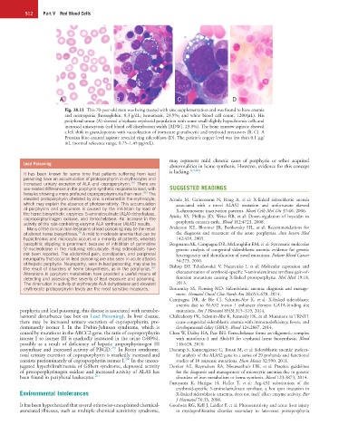
Fig. 38.11 This 70-year-old man was being treated with zinc supplementation and was found to have anemia
and neutropenia (hemoglobin, 8.3 g/dL; hematocrit, 23.9%; and white blood cell count, 1200/µL). His
peripheral smear (A) showed a biphasic erythroid population with some small slightly hypochromic cells and
increased anisocytosis (red blood cell distribution width [RDW], 23.9%). The bone marrow aspirate showed
a left shift in granulopoiesis with vacuolization of immature granulocytic and erythroid precursors (B, C). A
Prussian blue–stained aspirate revealed ring sideroblasts (D). The patient’s copper level was less than 0.1 µg/
mL (normal reference range, 0.75–1.45 µg/mL).
may represent mild chronic cases of porphyria or other acquired
Lead Poisoning
abnormalities in heme synthesis. However, evidence for this concept
It has been known for some time that patients suffering from lead is lacking. 263,264
poisoning have an accumulation of protoporphyrin in erythrocytes and
increased urinary excretion of ALA and coproporphyrin. 262 There are
sex-related differences in the porphyrin synthetic response to lead, with SUGGESTED READINGS
females showing a more profound coproporphyrinuria than men. 263 The
elevated protoporphyrin chelated by zinc is retained in the erythrocyte, Aivado M, Gattermann N, Rong A, et al: X-linked sideroblastic anemia
which may explain the absence of photosensitivity. This accumulation associated with a novel ALAS2 mutation and unfortunate skewed
of porphyrins and precursors is caused by the inhibition by lead of X-chromosome inactivation patterns. Blood Cells Mol Dis 37:40, 2006.
the heme biosynthetic enzymes: 5-aminolevulinate (ALA) dehydratase,
coproporphyrinogen oxidase, and ferrochelatase. An increase in the Ajioka RS, Phillips JD, Weiss RB, et al: Down-regulation of hepcidin in
activity of the rate-controlling enzyme ALA synthase (ALAS) results. porphyria cutanea tarda. Blood 112:4723, 2008.
Many of the clinical manifestations of lead poisoning may be the result Anderson KE, Bloomer JR, Bonkovsky HL, et al: Recommendations for
79
of altered heme biosynthesis. A mild to moderate anemia that can be the diagnosis and treatment of the acute porphyrias. Ann Intern Med
hypochromic and microcytic occurs in a minority of patients, whereas 142:439, 2005.
basophilic stippling is prominent because of inhibition of pyrimidine Bergmann AK, Campagna DR, McLoughlin EM, et al: Systematic molecular
5′-nucleotidase in the maturing reticulocyte. Ring sideroblasts have genetic analysis of congenital sideroblastic anemia: evidence for genetic
not been reported. The abdominal pain, constipation, and peripheral heterogeneity and identification of novel mutations. Pediatr Blood Cancer
neuropathy that occur in lead poisoning are also seen in acute attacks 54:273, 2010.
of hepatic porphyria. Neuropathy, seen in lead poisoning, may also be
the result of disorders of heme biosynthesis, as in the porphyrias. Bishop DF, Tchaikovskii V, Nazarenko I, et al: Molecular expression and
262
Alterations in porphyrin metabolism have provided a useful means of characterization of erythroid-specific 5-aminolevulinate synthase gain-of-
detecting and assessing the severity of lead exposure and poisoning. function mutations causing X-linked protoporphyria. Mol Med 19:18,
The diminution in activity of erythrocyte ALA dehydratase and elevated 2013.
erythrocyte protoporphyrin levels are the most sensitive measures. Bottomley SS, Fleming MD: Sideroblastic anemia: diagnosis and manage-
ment. Hematol Oncol Clin North Am 28:653–670, 2014.
Campagna DR, de Bie CI, Schmitz-Abe K, et al: X-linked sideroblastic
anemia due to ALAS2 intron 1 enhancer element GATA-binding site
porphyria and lead poisoning, this disease is associated with neurobe- mutations. Am J Hematol 89(3):315–319, 2014.
havioral disturbance (see box on Lead Poisoning). In liver disease, Chakraborty PK, Schmitz-Abe K, Kennedy EK, et al: Mutations in TRNT1
there may be increased urinary excretion of coproporphyrin, pre- cause congenital sideroblastic anemia with immunodeficiency, fevers, and
dominantly isomer I. In the Dubin-Johnson syndrome, which is developmental delay (SIFD). Blood 124:2867, 2014.
caused by mutations in the ABCC2 gene, the ratio of coproporphyrin Chen W, Dailey HA, Paw BH: Ferrochelatase forms an oligomeric complex
isomer I to isomer III is markedly increased in the urine (>80%), with mitoferrin-1 and Abcb10 for erythroid heme biosynthesis. Blood
possibly as a result of deficiency of hepatic uroporphyrinogen III 116:628, 2010.
259
cosynthase and increased activity of PBGD. In Rotor syndrome, Ducamp S, Kannengiesser C, Touati M, et al: Sideroblastic anemia: molecu-
total urinary excretion of coproporphyrin is markedly increased and lar analysis of the ALAS2 gene in a series of 29 probands and functional
260
consists predominantly of coproporphyrin isomer I. In the uncon- studies of 10 missense mutations. Hum Mutat 32:590, 2011.
jugated hyperbilirubinemia of Gilbert syndrome, depressed activity Donker AE, Raymakers RA, Nieuwenhuis HK, et al: Practice guidelines
of protoporphyrinogen oxidase and increased activity of ALAS has for the diagnosis and management of microcytic anemias due to genetic
been found in peripheral leukocytes. 261 disorders of iron metabolism or heme synthesis. Blood 123:3873, 2014.
Furuyama K, Harigae H, Heller T, et al: Arg-452 substitution of the
erythroid-specific 5-aminolaevulinate synthase, a hot spot mutation in
Environmental Intolerances X-linked sideroblastic anaemia, does not itself affect enzyme activity. Eur
J Haematol 76:33, 2006.
It has been hypothesized that several otherwise-unexplained chemical- Goodwin RG, Kell J, Laidler P, et al: Photosensitivity and acute liver injury
associated illnesses, such as multiple chemical sensitivity syndrome, in myeloproliferative disorder secondary to late-onset protoporphyria