
Page 593 - Hematology_ Basic Principles and Practice ( PDFDrive )
P. 593
508 Part V Red Blood Cells
rapid, with subnormal retention of the iron isotope in erythrocytes
1 after 10 to 14 days. Other features of ineffective erythropoiesis may
be variably present: a mild increase in bilirubin concentration,
decrease in haptoglobin levels, mild increase in lactate dehydrogenase
levels, and normal or slight increase in reticulocyte numbers. The
2 magnitude of iron overload correlates poorly with the degree of
anemia in patients who are not transfused. The degree of ineffective
erythropoiesis is a better predictor of the amount of iron overload.
When ferrokinetics are unavailable, the extent of erythroid hyperpla-
sia relative to normal acts as a rough measure of the magnitude of
3 ineffective erythropoiesis. Several studies have shown that the relative
increase in erythroid activity multiplied by the patient’s age shows a
good correlation with the degree of iron overload as measured by
plasma ferritin. 172,173 The iron overload does not result from muta-
174
tions in the HFE gene (see box on Therapy for Hereditary Sidero-
4 ? ? ? ? ? ? blastic Anemia).
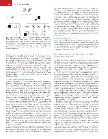
Fig. 38.9 PEDIGREE OF A FAMILY WITH PYRIDOXINE- Differential Diagnosis
RESPONSIVE SIDEROBLASTIC ANEMIA SHOWING X-LINKED Hereditary sideroblastic anemia should be distinguished from idio-
RECESSIVE INHERITANCE. Affected (filled box), carrier (filled circle pathic hemochromatosis, because both have biochemical evidence of
within open circle), and unknown status (question mark within circle or box) iron overload and a similar tissue pattern of iron deposition. Careful
47
are indicated. Diagonal lines indicate deceased members. This pedigree has hematologic assessment of patient and family members should make
been abbreviated to show only the affected branches of the family. The arrow the distinction, because the hemoglobin level and MCV are normal
indicates the proband.
in idiopathic hemochromatosis.
Other Nonsyndromic and Syndromic Hereditary
female carriers, although usually normal, can develop erythrocyte
dimorphism or varying degrees of anemia. The assignment of the Sideroblastic Anemias
158
gene for erythroid ALAS2 to the X chromosome and the many
mutations documented in erythroid ALAS2 provide the genetic basis X-linked sideroblastic anemia is considered the most common
for this X-linked disease. In several families, coinheritance of other inherited sideroblastic anemia; however, a number of rare forms have
X-linked traits (e.g., glucose-6-phosphate dehydrogenase [G6PD] recently been identified. These consist of two nonsyndromic sidero-
deficiency, ataxia with sideroblastic anemia) has been described. 168,169 blastic anemias, which have a similar phenotype to X-linked sidero-
Sporadic and familial cases have been described that affect only blastic anemia, and five syndromic forms where heme synthesis is
females, which has been shown to represent skewed X-chromosome affected in a variety of other tissues in addition to red cells.
inactivation (“unfortunate skewing”) affecting the normal allele for Of the nonsyndromic forms, inherited mutations in both the
170
the ALAS2 gene. The absence of affected male members in these SLC25A38 and GLRX5 genes have been identified to cause an
pedigrees suggests that the ALAS2 defects identified are lethal in autosomal recessive pyridoxine-refractory sideroblastic anemia. 178,179
hemizygous males. SLC25A38 is located on chromosome 3p22.1 and encodes a mito-
chondrial carrier protein that may function to import glycine into
Clinical and Laboratory Evaluation the mitochondrion or exchange glycine for 5-aminolevulinate.
179
Typically the anemia of X-linked sideroblastic anemia manifests in Homozygous or compound heterozygote mutations in SLC25A38
infancy or childhood, but the milder forms of anemia may not be result in a similar phenotype to that seen in X-linked sideroblastic
found until midlife. Even elderly patients have been diagnosed with anemia, with onset in infancy of a severe microcytic anemia that is
171
161
this anemia. Some cases may be discovered only during family refractory to treatment with pyridoxine and folic acid. GLRX5
surveys, which should always be undertaken when hereditary sidero- encodes a mitochondrial protein, glutaredoxin 5, which when deleted
blastic anemia is diagnosed. Still other patients may present with in the zebrafish mutant shiraz results in defective iron-sulfur cluster
180
features of iron overload, such as diabetes or cardiac failure. Iron assembly and blocked synthesis of heme. A late-onset pyridoxine-
overload occurs commonly even with mild anemia and may occasion- refractory sideroblastic anemia caused by homozygous mutation in
ally be seen with female carriers. Enlargement of the liver and spleen GLRX5 has been described in a patient who in middle age developed
may occur with mild abnormalities of liver function tests. symptoms of a microcytic hypochromic anemia, type 2 diabetes,
Anemia is extremely variable, but even when little or no anemia cirrhosis, and liver iron overload. 178
is present, the mean corpuscular volume (MCV) is low, and the red In addition to genetically defined forms of hereditary sideroblastic
cell volume distribution width may be increased. When anemia is anemia, five syndromic types have been described, which present with
3
severe, the MCV may be as low as 50 fL (50 µm ). The blood smear anemia in combination with either muscle, neurologic, or pancreatic
shows a population of cells with hypochromic, microcytic morphol- tissue involvement. The first of these disorders to be defined by
ogy (see Fig. 38.8), which contrasts with the other normochromic, molecular genetics, the Pearson syndrome, is a rare entity that mani-
normocytic cells (i.e., dimorphism). Anisocytosis, poikilocytosis, fests in early infancy with anemia and exocrine pancreatic dysfunc-
181
elongated cells, and siderocytes may also be seen. The characteristic tion. The anemia is normocytic or macrocytic, reticulocyte counts
erythrocyte dimorphism is most prominent in patients with milder are low, and variable degrees of neutropenia and thrombocytopenia
anemia, in female carriers, and in patients in whom pyridoxine has are present. The bone marrow shows striking vacuolation and ringed
182
corrected the anemia but not restored the MCV to normal. In some sideroblasts. Although usually fatal, milder forms of the anemia are
pedigrees with only affected females, macrocytosis may be present, consistent with survival into adult life. The syndrome, which is
which contrasts with the typical microcytosis of male hemizy- related to the Kearns-Sayre syndrome, results from deletions, muta-
gotes. 141,170 Leukocyte values are normal, whereas the platelet count tions, or duplications of mitochondrial DNA, variably affecting
is normal or increased. multiple tissues of the body. 183,184
Serum iron concentration is increased, and transferrin shows an A second syndromic congenital sideroblastic anemia, X-linked
increased percentage of saturation with iron. Serum ferritin levels are sideroblastic anemia with cerebellar ataxia (XLSA/A), is a rare mito-
invariably increased. Ineffective erythropoiesis can be confirmed by chondrial disease caused by loss-of-function mutations in the ATP-
ferrokinetic measurements showing that plasma iron clearance is binding cassette transporter ABCB7. 185–188 ABCB7 is localized to the