
Page 688 - Hematology_ Basic Principles and Practice ( PDFDrive )
P. 688
580 Part V Red Blood Cells
HbS RBC responses. As one example, the growth factor angiopoietin-2, released
polymer sickling from Weibel-Palade bodies, sensitizes the endothelial cell toward
↓pO 2
↑[HbS] deoxyHbS Vascular TNF-α and consequent adhesion molecule expression. It is not pos-
+ Hemolysis Stasis sible to identify proportionate contributions of the many specific
inflammatory inputs, but ligand-triggered TLR4 signaling is emerg-
ing as an important contributor. 17
↑ RBC & WBC adhesion Free heme & In summation, sickle cell anemia is a chronic, systemic inflamma-
19
to endothelium hemoglobin tory state. Characteristic features include: leukocytosis; activation
Vascular of granulocytes, monocytes, lymphocyte subsets, and mast cells;
Occlusion elevated levels of proximate inflammatory mediators, acute-phase
reactants, and distal effectors; abundance of soluble adhesion mol-
Systemic ecules; activation of coagulation; presence of microparticles from
Endothelial Systemic multiple activated cell types; and generation of excess oxidant via
Dysfunction Inflammation Ischemia multiple mechanisms. Importantly, this systemic inflammatory state
reperfusion is perpetual, with footprints of active inflammation apparent even
between acute clinical events. Clinically, leukocytosis in sickle cell
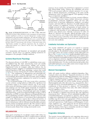
Fig. 41.10 ISCHEMIA/REPERFUSION AS THE CORE DRIVING disease is a risk factor for mortality, clinical and silent stroke, and
FORCE IN SICKLE CELL ANEMIA. As the consequence of vasoocclusion, acute chest syndrome, and it helps predict which babies will develop
ischemia–reperfusion provides an incessant driving force causing systemic a severe clinical course. Conversely, several clinical complications are
inflammation with microvascular dysfunction. The adhesion biology result- themselves overtly inflammatory, in particular acute painful episodes
ing from activated/dysfunctional endothelial cells creates a positive feedback and acute chest syndrome.
loop that slows microvascular transit and enables polymer formation. (Modi-
fied from Hebbel RP: Reconstructing sickle cell disease: A data-based analysis of the
“hyperhemolysis paradigm” for pulmonary hypertension from the perspective of Endothelial Activation and Dysfunction
evidence-based medicine. Am J Hematol 86:123, 2011.) Hb, Hemoglobin; PS,
phosphatidylserine.
The vascular endothelial cell receives and responds to disparate
inputs, both sensing and modifying its environment. Although
First, vasoocclusion and hemolysis are interrelated and mutually normally adaptive, the endothelial response can become maladaptive.
promotive. Second, there is an apparent unifying explanation for In the sickle context, the extreme complexity of the sickle milieu (see
emergence of this panoply of biologic mediators. Box on The Complex Sickle Milieu) molds endothelial cell function,
leading to a harmful state. The core inflammatory and oxidative input
causes a high level of endothelial activation with adverse consequences
Ischemia-Reperfusion Physiology such as coagulation activation, degradation of the endothelial glyco-
calyx (a critical determinant of endothelial cell homeostasis), and
The abnormal adhesion of sickle RBC to endothelium creates a posi- endothelial dysfunction. An often-overlooked principle predicts that
tive feedback loop that slows microvascular transit and thereby endothelial dysfunction in the sickle context creates unique risk (see
enables deoxygenation, polymerization, sickling, and occlusion. Mortality and Sudden Death, later).
Experimental studies in transgenic sickle mice indicate that the
enabling, proximate instigation of endothelial activation derives from
ischemia-reperfusion (I/R) injury physiology, a process that would Aberrant Vasoregulation
comprise an incessant driving force for systemic inflammation (Fig.
18
41.10). This combination of inflammation and endothelial dys- Sickle cell anemia involves deficient endothelial-dependent, flow-
20
function can provide a unifying explanation for the multitude of mediated conduit artery dilation. Beyond the dominant role of I/R
vascular biologic abnormalities in sickle cell anemia, and it establishes and inflammation-induced endothelial dysfunction, there are several
a contextual and generative explanation for several of the specific additional contributing processes: TNF-α induction of endothelial
clinical complications. L-arginase, limiting L-arginine availability to eNOS; NO consump-
The complicated biology of I/R injury occurs when a proximate tion by excess superoxide and cell-free oxyHb; elevated asymmetric
vascular occlusion causing ischemia is followed by reperfusion that dimethylarginine; plus other aberrancies (e.g., microparticles, abnor-
12
reintroduces oxygen to the formerly ischemic area. In the unique mal wall shear stress, elevated phospholipase A2). The proportionate
sickle context, the initiating occlusive event(s) would be microvascular contributions of these processes is unknown. NO bioavailability
and multifocal, and they would happen recurrently. As revealed by seems to be higher for females than males, who exhibit substantial
studies of sickle transgenic mice, this triggers the classic, reperfusion- non-NO regulation of flow.
dependent, early I/R events of localized and rapid onset of oxygen Perfusion patterns are complex in sickle cell anemia but can gener-
radical generation, nuclear factor kappa-B (NFκB) activation, TNF-α ally be described as impaired microvascular flow and augmented
21
production, and WBC activation. Research on I/R generally illustrates macrovascular flow. Aberrancy of both endothelial-dependent and
that its pathobiology thereafter explosively arborizes to become vastly endothelial-independent vasoregulation is apparent, and the milieu
complex. Its hallmark, however, is conversion of this localized process is replete with a multitude of vasoactive substances. A state of vascular
into a sterile inflammatory response that is robust and systemic. It instability may derive from tonic upregulation of both vasoconstric-
initiates widespread microvascular dysfunction and organ accumula- tive and vasodilatory systems, in addition to exaggerated α 1 -adrenergic
tions of inflammatory cells by infiltration from blood plus activation vessel wall responsiveness. Sickle humans reveal disruption of auto-
of tissue resident macrophages and mast cells. This can lead to disease nomic regulation (e.g., with augmented risk for hypoxia-induced
22
remote from the initiating occlusion, and it can explain emergence perfusion decrements). Systemic blood pressure in patients with
of macrovascular disease from inciting microvascular events. sickle cell anemia is lower than in nonanemic controls, yet is higher
than in comparably anemic β-thalassemics.
INFLAMMATION
Coagulation Activation
Many agents comprising the exceedingly complex sickle milieu (see
Pink Box) exert proinflammatory effects; a number interact with each The interplay between inflammation and coagulation often seen in
other in ways that alter the nature or complexity or magnitude of biomedicine is generally highly evident in sickle cell anemia, with