
Page 693 - Hematology_ Basic Principles and Practice ( PDFDrive )
P. 693
Chapter 42 Sickle Cell Disease 585
D
A B C E
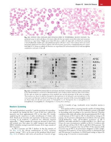
Fig. 42.1 SICKLE CELL DISEASE AND HEMOGLOBIN SC PERIPHERAL BLOOD SMEARS. The
peripheral smear in sickle cell disease (A) shows sickle cells that are mostly irreversibly sickled and sometimes
referred to as “cigar forms.” Higher power detail (B) shows a sickle cell (upper left), red blood cell containing
a Howell-Jolly body (middle right), and polychromatophilic cell (lower center). These indicate sickle cell anemia
and splenic dysfunction but marrow response with reticulocytosis, respectively. A peripheral smear of a patient
with Hgb SC (C) shows no sickled cell, but there are target forms (D) and occasional cells (E) with hemoglobin
condensed at each pole of the cell.
Fig. 42.2 COMPARATIVE ANALYSES OF SEVERAL MUTANT HEMOGLOBINS USING ALKALINE
ELECTROPHORESIS, ACID ELECTROPHORESIS, AND THIN-LAYER ISOELECTRIC FOCUSING.
On the right are shown the components of the standard (top) and the phenotypes of the other six samples.
Their analyses are shown by alkaline hemoglobin electrophoresis in the left panel, acid electrophoresis in the
center panel, and thin-layer isoelectric focusing in the right panel. Locations of the various hemoglobin bands
are shown below the left and center panels. A, Hemoglobin A; A2, hemoglobin A2; C, hemoglobin C; E,
hemoglobin E; F, hemoglobin F; S, hemoglobin S. (Courtesy M.H. Steinberg.)
and by 4 months of age, moderately severe hemolytic anemia is
Newborn Screening evident.
Tests used in newborn screening must be capable of distinguishing
10
The use of prophylactic penicillin and the provision of comprehen- between Hb F, S, A, and C. The Hb distribution pattern is described
sive medical care during the first 5 years of life have reduced the in descending order according to the quantities detected. Therefore
mortality rate from approximately 25% to less than 3%, thereby a newborn with sickle cell anemia who has predominantly Hb F with
underlining the importance of early identification of infants with a small amount of Hb S and no Hb A is described as having an FS
SCD. Based on its economy and superiority of detection, universal pattern. An FS pattern is obtained also in newborns who have sickle
screening of all newborns is preferred over ethnically targeted cell–β°-thalassemia, sickle cell–hereditary persistence of Hb F
approaches. 11,12 Blood samples for testing are obtained by heel stick (HPFH), and sickle cell–Hb D or sickle cell–Hb G (i.e., Hb D and
and spotted onto filter paper for stable transport and subsequent E have the same electrophoretic mobility as Hb S). A newborn with
HPLC (solubility testing is unreliable because of the large amount of sickle cell trait will have Hb F, Hb A, and Hb S (FAS pattern). The
Hb F present.) quantity of Hb A is greater than that of Hb S. If the quantity of Hb
+
As Hb S increases and Hb F declines in the first months of S exceeds that of Hb A, the presumptive diagnosis is sickle cell–β -
life (Fig. 42.3), the clinical manifestations of SCD, including thalassemia (FSA pattern). It may not be possible to distinguish FAS
13
anemia, emerge. ISCs can be seen on the peripheral blood smear and FSA patterns in newborns, so DNA-based testing or repeat Hb
(Fig. 42.4) of children with sickle cell anemia at 3 months of age, testing at age 3–6 months is recommended.