
Page 74 - Hematology_ Basic Principles and Practice ( PDFDrive )
P. 74
46 Part I Molecular and Cellular Basis of Hematology
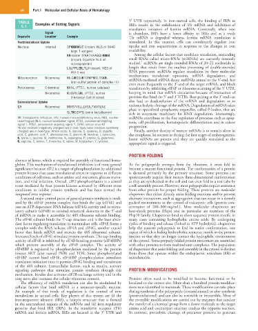
TABLE Examples of Sorting Signals 5′ UTR respectively. In iron-starved cells, the binding of IRPs to
5.1 IREs results in the stabilization of Tfr mRNA and inhibition of
translation initiation of ferritin mRNA. Conversely, when iron
Signal is abundant, IRPs have a lower affinity to IREs and as a result
Organelle Location a Example Tfr mRNA is degraded whereas ferritin mRNA translation is
Posttranslational Uptake stimulated. In this manner, cells can coordinately regulate iron
Nucleus Internal SPKKKRKVE (import; NLS of SV40 uptake and iron sequestration in response to the changes in iron
large T antigen) availability.
KR-spacer (PAATKKAGQ)-KKKK Among the cellular factors that modulate translation, noncoding
(import; bipartite NLS of small RNAs called micro-RNAs (miRNAs) are currently intensely
1
nucleoplasmin) studied. miRNAs are single stranded RNAs of 20–22 nucleotide in
LQLPPLERLTLD (export; NES of length that result from the nuclear processing of double-stranded
HIV-1 rev) RNA precursors. miRNAs regulate translation by three molecular
mechanisms: translation repression, mRNA degradation, and
Mitochondrion N-terminal MLGIRSSVKTCFKPMSLTSKRL miRNA-mediated mRNA decay. miRNAs anneal to the 5′-end, but
(iron-sulfur protein of complex III)
even more frequently to the 3′-end of the target mRNA, and block
Peroxisomes C-terminal KANL (PTS1, human catalase) translation by inhibiting eIF4F or ribosome scanning of the 5′ UTR,
N-terminal RLQVVLGHL (PTS2, human keeping in mind that mRNA circularizes because of interaction of
3-ketoacyl-CoA thiolase) proteins that bind the 5′ and 3′ UTRs. Base pairing at the 3′-end can
Cotranslational Uptake also lead to deadenylation of the mRNA and degradation or to
ER N-terminal MMSFVSLLLVGILFWATEAE endonucleolytic cleavage of the mRNA. Degradation of mRNA takes
place in specialized cytoplasmic organelles, called P-bodies, that are
QLTKCEVFQ (ovine lactalbumin) rich in enzymatic machinery for RNA degradation. Interestingly,
ER, Endoplasmic reticulum; HIV, human immunodeficiency virus; NES, nuclear miRNAs contribute to the fine regulation of processes such as apop-
export signal; NLS, nuclear localization signal; PTS1, peroxisomal targeting tosis, cell proliferation, hematopoietic differentiation and in cancer
signal-1; PTS2, peroxisomal targeting signal-2; SV40, simian virus 40.
a Acidic residues (negatively charged) are in italic type; basic residues (positively progression.
charged) are in bold type. Amino acids: A, alanine; C, cysteine; D, aspartic Finally, another destiny of mature mRNAs is to remain silent in
acid; E, glutamic acid; F, phenylalanine; G, glycine; H, histidine; I, isoleucine; the cytoplasm. In oocytes or during the first stages of embryogenesis,
K, lysine; L, leucine; M, methionine; N, asparagine; P, proline; Q, glutamine; latent mRNAs are present and they are quickly translated as the
R, arginine; S, serine; T, threonine; V, valine; W, tryptophan; Y, tyrosine.
appropriate signal is triggered.
PROTEIN FOLDING
absence of heme, which is required for assembly of functional hemo-
globin. This mechanism of translational inhibition is of more general As the polypeptide emerges from the ribosome, it must fold to
significance because eIF2 is a target of phosphorylation by additional become a mature functional protein. The conformation of a protein
protein kinases that cause translational arrest in response to different is dictated primarily by the primary structure. Some proteins can
conditions of cell stress, such as amino acid starvation, glucose starva- spontaneously acquire their mature three-dimensional conformation
tion, and viral infection. Overall, eIF2 phosphorylation is a central as they are synthesized in the cell and can even fold in a test tube by
event mediated by four protein kinases activated by different stress a self-assembly process. However, most polypeptides require assistance
conditions to inhibit protein synthesis and has been termed the from other protein for proper folding. These proteins are molecular
integrated stress response. chaperones that either directly assist folding reactions and/or prevent
A second major control point of general protein synthesis is medi- aberrant interactions, such as aggregation that can occur in a densely
ated by the eIF4F protein complex that binds the cap (eIF4E) and packed environment as the cytosol of eukaryotic cells (protein con-
uses an ATP-dependent RNA helicase (eIF4A) activity and its stimu- centration of 200–300 mg/mL). Most molecular chaperones are
latory subunit (eIF4B) to unwind structural elements in the 5′-end heat-shock proteins (Hsps) and in particular are members of the
of mRNA to make it accessible for 40S ribosome subunit binding. Hsp70 family. Chaperones bind to short sequence protein motifs, in
The eIF4E subunit binds the 5′-cap structure and is the least abun- many cases containing hydrophobic amino acids. By undergoing
dant factor regulating translation in mammalian cells. eIF4E forms a cycles of binding and release (linked to ATP hydrolysis), chaperones
complex with the RNA helicase eIF4A and eIF4G, another crucial help the nascent polypeptide to find its native conformation, one
factor that binds mRNA and recruits the 40S ribosomal subunit. aspect of which is hiding hydrophobic sequence motifs in the protein
Increased levels of eIF4E stimulate protein synthesis. The cap-binding interior so that they no longer contact the hydrophilic environment
activity of eIF4E is inhibited by eIF4E-binding proteins (eIF4EBPs) of the cytosol. Some properly folded protein monomers are assembled
which prevent assembly of the eIF4F complex. The activity of with other proteins to form multisubunit complexes. The population
eIF4EBP is regulated by phosphorylation mediated by the protein of chaperones that assist folding and assembly in the cytosol is distinct
kinases AKT (also named PKB) and TOR. Since phosphorylated from those that operate within the endoplasmic reticulum (ER) or
eIF4BP cannot bind eIF4E, eIF4EBP phosphorylation stimulates mitochondria.
translation initiation since it permits eIF4G binding and recruitment
of the 40S subunit. Extracellular factors, such as insulin, activate
signaling pathways that stimulate protein synthesis through this PROTEIN MODIFICATIONS
mechanism. Insulin also activates eIF2B exchange activity and in the
long term also increases the cellular ribosome content. Proteins often need to be modified to become functional or be
The efficiency of mRNA translation can also be modulated by localized to the correct site. More than a hundred protein modifica-
cellular factors that bind mRNA in a sequence-specific manner. tions were identified in mammals. These modifications can take place
An example of this mode of regulation is the control of iron during synthesis of the polypeptide (cotranslational) or after synthesis
metabolism in animal cells. Key players of this system are (i) the (posttranslational) and can also be reversible or irreversible. Most of
iron-responsive element (IRE), a hairpin structure that is formed the reversible modifications are carried out by enzymes that catalyze
in the untranslated regions of the mRNAs and (ii) iron regulatory the transfer of a chemical group from a donor molecule to the target
proteins that bind IRE (IRPs). In the transferrin receptor (Tfr) amino acid and counterpart enzymes catalyze the opposite reaction.
mRNA and ferritin mRNA, IREs are located in the 3′ UTR and In contrast, proteolytic cleavage of precursor proteins to generate