
Page 763 - Hematology_ Basic Principles and Practice ( PDFDrive )
P. 763
650 Part V Red Blood Cells
T-cell deficiencies) T-cell and humoral immune deficiency predispose female <12.0 g/dL), reticulocytosis (corrected reticulocyte count
to WAIHA, but no correlation between the type and severity of >2% or absolute reticulocyte count >100,000/µL to 120,000/µL),
immune deficiency and the risk of AIHA has been established. One low haptoglobin, elevated lactate dehydrogenase (LDH), and elevated
phenomenon that is poorly understood is the lack of a clear relation- unconjugated (indirect) bilirubin. Haptoglobin is an α2-globulin
ship between the presence of RBC antibodies and anemia. In many that binds Hb. This Hb–haptoglobin complex is degraded in the
instances, there is no anemia despite a strongly positive DAT or high liver. Hemopexin is another plasma protein with a very high binding
titers of CAIHAs. There is also only a poor correlation between affinity to Hb. It scavenges heme released from RBCs and protects
antibody titers and severity of anemia. Another unexplained finding the organisms from the adverse effects of circulating Hb. The deter-
in secondary AIHA is the occurrence of both WAIHAs and CAIHAs mination of hemopexin is not essential for the diagnosis of AIHA.
in the some condition; for example, in lymphomas or infections. Indirect bilirubin is usually not more than 5 mg/dL except in associ-
Antibodies in primary AIHA are frequently polyreactive and ated liver disease (Epstein-Barr [EBV]-associated AIHA). Additional
polyclonal (no clonal B cells detected by polymerase chain reaction findings are increased urobilinogen in the urine and spherocytes in
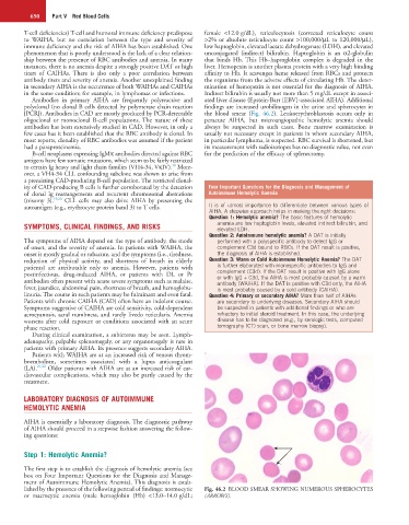
[PCR]). Antibodies in CAD are mostly produced by PCR-detectable the blood smear (Fig. 46.2). Leukoerythroblastosis occurs only in
oligoclonal or monoclonal B-cell populations. The nature of these peracute AIHA, but microangiopathic hemolytic anemia should
antibodies has been extensively studied in CAD. However, in only a always be suspected in such cases. Bone marrow examination is
few cases has it been established that the RBC antibody is clonal. In usually not necessary except in patients in whom secondary AIHA,
most reports, clonality of RBC antibodies was assumed if the patient in particular lymphoma, is suspected. RBC survival is shortened, but
had a paraproteinemia. its measurement with radioisotopes has no diagnostic value, not even
B-cell neoplasms expressing IgMκ antibodies directed against RBC for the prediction of the efficacy of splenectomy.
antigens have few somatic mutations, which seem to be fairly restricted
18
to certain Ig heavy and light chain families (VH4-34, VκIV). More-
over, a VH4-34 CLL confounding subclone was shown to arise from
a preexisting CAD-producing B-cell population. The restricted clonal-
ity of CAD-producing B cells is further corroborated by the detection Four Important Questions for the Diagnosis and Management of
of clonal Ig rearrangements and recurrent chromosomal aberrations Autoimmune Hemolytic Anemia
(trisomy 3). 19,20 CLL cells may also drive AIHA by presenting the
autoantigen (e.g., erythrocyte protein band 3) to T cells. It is of utmost importance to differentiate between various types of
AIHA. A stepwise approach helps in making the right decisions:
Question 1: Hemolytic anemia? The basic features of hemolytic
SYMPTOMS, CLINICAL FINDINGS, AND RISKS anemia are low haptoglobin levels, elevated indirect bilirubin, and
elevated LDH.
Question 2: Autoimmune hemolytic anemia? A DAT is initially
The symptoms of AIHA depend on the type of antibody, the mode performed with a polyspecific antibody to detect IgG or
of onset, and the severity of anemia. In patients with WAIHA, the complement C3d bound to RBCs. If the DAT result is positive,
onset is mostly gradual or subacute, and the symptoms (i.e., tiredness, the diagnosis of AIHA is established.
reduction of physical activity, and shortness of breath in elderly Question 3: Warm or Cold Autoimmune Hemolytic Anemia? The DAT
patients) are attributable only to anemia. However, patients with is further elaborated with monospecific antibodies to IgG and
postinfectious, drug-induced AIHA, or patients with DL or Pr complement (C3d). If the DAT result is positive with IgG alone
or with IgG + C3d, the AIHA is most probably caused by a warm
antibodies often present with acute severe symptoms such as malaise, antibody (WAIHA). If the DAT is positive with C3d only, the AIHA
fever, jaundice, abdominal pain, shortness of breath, and hemoglobu- is most probably caused by a cold antibody (CAIHA).
linuria. The course in such patients may be fulminant and even fatal. Question 4: Primary or secondary AIHA? More than half of AIHAs
Patients with chronic CAIHA (CAD) often have an indolent course. are secondary to underlying diseases. Secondary AIHA should
Symptoms suggestive of CAIHA are cold sensitivity, cold-dependent be suspected in patients with additional findings or who are
acrocyanosis, acral numbness, and rarely livedo reticularis. Anemia refractory to initial steroid treatment. In this case, the underlying
worsens after cold exposure or conditions associated with an acute disease has to be diagnosed (e.g., by serologic tests, computed
phase reaction. tomography (CT) scan, or bone marrow biopsy).
During clinical examination, a subicterus may be seen. Lymph-
adenopathy, palpable splenomegaly, or any organomegaly is rare in
patients with primary AIHA. Its presence suggests secondary AIHA.
Patients with WAIHA are at an increased risk of venous throm-
boembolism, sometimes associated with a lupus anticoagulant
(LA). 21,22 Older patients with AIHA are at an increased risk of car-
diovascular complications, which may also be partly caused by the
treatment.
LABORATORY DIAGNOSIS OF AUTOIMMUNE
HEMOLYTIC ANEMIA
AIHA is essentially a laboratory diagnosis. The diagnostic pathway
of AIHA should proceed in a stepwise fashion answering the follow-
ing questions:
Step 1: Hemolytic Anemia?
The first step is to establish the diagnosis of hemolytic anemia (see
box on Four Important Questions for the Diagnosis and Manage-
ment of Autoimmune Hemolytic Anemia). This diagnosis is estab-
lished by the presence of the following pentad of findings: normocytic Fig. 46.2 BLOOD SMEAR SHOWING NUMEROUS SPHEROCYTES
or macrocytic anemia (male hemoglobin (Hb) <13.0–14.0 g/dL; (ARROWS).