
Page 766 - Hematology_ Basic Principles and Practice ( PDFDrive )
P. 766
Chapter 46 Autoimmune Hemolytic Anemia 653
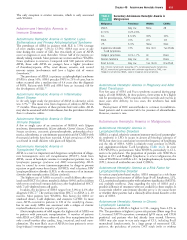
The only exception is ovarian teratoma, which is only associated TABLE Secondary Autoimmune Hemolytic Anemia in
with WAIHA. 46.2 Malignancies
Malignancy Prevalence WAIHAs CAIHAs
Autoimmune Hemolytic Anemia in MGUS Very low None All
Immune Diseases All NHL 0.23–2.6%
CLL 4.3–9% 90% 10%
Autoimmune Hemolytic Anemia in Systemic Lupus SMZL 10% 2/3 1/3
Erythematosus and Primary Antiphospholipid Syndrome
The prevalence of AIHA in patients with SLE is 7.5% (average LPL 3–5% None Most
of seven studies; range: 5.2% to 12.5%). AIHA may occur at any Angioimmunoblastic 13% One third Two thirds
time during the course of SLE, but two-thirds of cases of AIHA T-cell lymphoma
occur at diagnosis or soon thereafter. Almost half of the patients are Hodgkin lymphoma 0.19–1.7% All None
already taking steroids. Most patients are women and have WAIHA. Ovarian teratoma Very low All None
Evans syndrome is common. Compared with SLE patients without
AIHA, those with AIHA are younger; have a higher prevalence Solid tumors Very low Two thirds One third
of thrombocytopenia, APAs, renal disease, serositis, and central CLL, Chronic lymphocytic leukemia; LPL, lymphoplasmacytic lymphoma;
nervous system involvement; and have a higher risk of venous MGUS, monoclonal gammopathy with unknown significance; NHL, non-
thrombosis. 27 Hodgkin’s lymphoma; SMZL, splenic marginal zone lymphoma.
The prevalence of AIHA in primary antiphospholipid syndrome
(PAPS) is about 10%. AIHA precedes PAPS in 25% of cases, but in
others occurred after a median time of 4.4 years after the diagnosis
of PAPS. Patients with PAPS and AIHA have an increased risk for Autoimmune Hemolytic Anemia in Pregnancy and After
the development of SLE. 28 Blood Transfusion
Very few cases of AIHA and Evans syndrome occurred during preg-
Autoimmune Hemolytic Anemia in Inflammatory nancy, all with WAIHAs. In these patients, there seems to be a higher
Bowel Disease risk for preeclampsia. AIHA responds well to steroids and resolves in
In the only larger study the prevalence of AIHA in ulcerative colitis most cases after delivery. In two cases, the newborns had mild
28a
was 1.7%. The mean time from diagnosis of colitis to AIHA was hemolysis.
17 months. Three-quarters of these patients had total colitis. AIHA Development of RBC autoantibodies is common in multitrans-
may also be associated with Crohn disease, but the prevalence is lower fused patients and is associated with the presence of alloantibodies.
than in ulcerative colitis. However, anemia is rare.
Autoimmune Hemolytic Anemia in Other
Immune Diseases Autoimmune Hemolytic Anemia in Malignancies
A few or single cases of an association of WAIHA with Sjögren
syndrome, dermatomyositis, biliary cirrhosis, Graves’ disease, Churg- Autoimmune Hemolytic Anemia in
Strauss syndrome, crescentic glomerulonephritis, polymyalgia rheu- Lymphoproliferative Disorders
matica, scleroderma, or autoimmune pancreatitis and of CAIHA with AIHA is a typical, relatively common immune-mediated paraneoplas-
rheumatoid arthritis have been reported. A high prevalence of AIHA tic syndrome in LPD. It occurs in almost all histologic subtypes of
was found in small children with giant-cell hepatitis. NHL, but there is no correlation between the frequency of lymphoma
and the risk of AIHA. AIHA is relatively most common in SMZL
Autoimmune Hemolytic Anemia in and angioimmunoblastic T-cell lymphoma (Table 46.2). In most
Transplanted Patients LPD WAIHAs is predominant. Most WAIHAs, particularly in CLL,
AIHA is a rare but important and dangerous complication of alloge- seem to be polyclonal. The proportion of patients with WAIHAs is
neic hematopoietic stem cell transplantation (HSCT). Aside from highest in CLL and Hodgkin lymphoma; in other malignancies, the
AIHA, causes of hemolytic anemia in transplanted patients may be ratio of WAIHAs to CAIHAs is 2:1. In lymphoplasmacytic lymphoma
lymphocyte passenger syndrome and ABO incompatibility. AIHA (LPL), almost all antibodies are clonal CAIHAs.
may be caused by severe immunosuppression by drugs to prevent
rejection or graft-versus-host disease (GVHD), viral infections, EBV Autoimmune Hemolytic Anemia as a Risk Factor for
lymphoproliferative disorder (LPD), or the recurrence of an immune Lymphoproliferative Disorder
disorder after transplantation (biliary cirrhosis). In various population-based studies, AIHA emerged as a risk factor
The highest rate of AIHA occurred in small children after unre- for subsequent development of diffuse large B-cell lymphoma, LPL,
lated cord HSCT for inborn metabolic defects (44%) and in children CLL, monoclonal gammopathy with unknown significance (MGUS),
with severe combined immune deficiency after haploidentical HSCT and multiple myeloma. Data regarding the type of AIHA and a
with T-cell–depleted stem cell grafts. possible influence of treatment were not available in these studies. It
In adults, the incidence of AIHA ranges from 3.0% to 4.4% after is uncertain whether autoimmune disorder per se is the causal factor
29
allogeneic HSCT. The median time from HSCT to AIHA is 4–10 or whether these patients had a clinically silent clonal disorder at the
months. Most patients have WAIHAs. Risk factors for AIHA are time of AIHA.
unrelated donor, T-cell depletion, and extensive GVHD. In most
cases, AIHA occurred in patients in CR of the underlying disease, Autoimmune Hemolytic Anemia in Chronic
but in one study AIHA was associated with a relapse of chronic Lymphocytic Leukemia
myeloid leukemia in the majority of patients. The prevalence of AIHA is highest in CLL, ranging from 4.3% to
After transplantation of solid organs, the highest risk of AIHA was 9%. The prevalence is highest in poor-risk patients (Binet stage B or
in patients with pancreatic transplantation. A number of patients C, increased ZAP70 expression, unmutated IgVH status, and CD38
with AIHA or CAIHA were observed after liver transplantation but positivity) and patients who had already been treated. However,
only a small number after cardiac, lung, intestinal, and renal trans- AIHA may also occur in very early stages of CLL, including B-cell
plantation. The most likely cause of AIHA in these patients is severe monoclonal lymphocytosis. In a large group of nontreated CLL
drug-induced immunosuppression. patients, the prevalence of positive DAT result (with or without