
Page 817 - Hematology_ Basic Principles and Practice ( PDFDrive )
P. 817
Chapter 50 Disorders of Phagocyte Function 703
has been identified have point mutations in a stretch of 250 amino C3b CD11b
acids in the extracellular domain of CD18. This region is highly
conserved among all β subunits and appears to be important for
Normal 100%
interaction with the α subunit.
Clinical Features
The key features of LAD I are summarized in Table 50.7. The clinical
presentation of LAD is heterogeneous and is related to the severity 0.1%
of the deficiency of the β2 integrins. The severe clinical phenotype Severe
is associated with less than 0.3% of the normal amount of these
glycoproteins on the leukocyte surface; the moderate phenotype has
2.5– 6% of normal levels. In both the severe and moderate forms of Cell number
the disease, persistent granulocytosis (neutrophil count of 12,000– 4.0%
3
100,000/mm ) is a constant finding, as are recurrent cutaneous
abscesses and aggressive periodontitis and gingivitis. Additional clini- Moderate
cal features seen more often in the severe clinical phenotype include
delayed umbilical cord separation, omphalitis, perirectal cellulitis,
severe ulcerative stomatitis, and bacterial sepsis. A striking finding in
LAD I is that abscesses and other sites of infections are devoid of pus 60%
despite the marked neutrophilia because neutrophils are unable to Hetero
emigrate to tissues. S. aureus and gram-negative enteric bacteria cause
the majority of infections in LAD I. Fungal infections can also occur,
particularly from C. albicans and Aspergillus spp.
Note that infants with delayed separation of the umbilical cord Log fluorescence intensity
who are healthy and have normal blood counts are very unlikely to
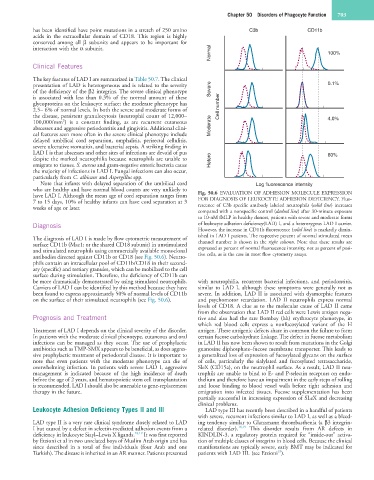
have LAD I. Although the mean age of cord separation ranges from Fig. 50.6 EVALUATION OF ADHESION MOLECULE EXPRESSION
7 to 15 days, 10% of healthy infants can have cord separation at 3 FOR DIAGNOSIS OF LEUKOCYTE ADHESION DEFICIENCY. Fluo-
weeks of age or later. rescence of C3b specific antibody labeled neutrophils (solid line) increases
compared with a nonspecific control (dashed line) after 30-minute exposure
to 10-nM fMLP in healthy donors, patients with severe and moderate forms
Diagnosis of leukocyte adhesion deficiency(LAD) I, and a heterozygous LAD I carrier.
However, the increase in CD11b fluorescence (solid line) is markedly dimin-
ished in LAD I patients. The respective percent of normal stimulated mean
The diagnosis of LAD I is made by flow cytometric measurement of
surface CD11b (Mac1; or the shared CD18 subunit) in unstimulated channel number is shown in the right column. Note that these results are
and stimulated neutrophils using commercially available monoclonal expressed as percent of normal fluorescence intensity, not as percent of posi-
antibodies directed against CD11b or CD18 (see Fig. 50.6). Neutro- tive cells, as is the case in most flow cytometry assays.
phils contain an intracellular pool of CD11b/CD18 in their second-
ary (specific) and tertiary granules, which can be mobilized to the cell
surface during stimulation. Therefore, the deficiency of CD11b can
be more dramatically demonstrated by using stimulated neutrophils. with neutrophilia, recurrent bacterial infections, and periodontitis,
Carriers of LAD I can be identified by this method because they have similar to LAD I, although these symptoms were generally not as
been found to express approximately 50% of normal levels of CD11b severe. In addition, LAD II is associated with dysmorphic features
on the surface of their stimulated neutrophils (see Fig. 50.6). and psychomotor retardation. LAD II neutrophils express normal
levels of CD18. A clue as to the molecular cause of LAD II came
from the observation that LAD II red cells were Lewis antigen nega-
Prognosis and Treatment tive and also had the rare Bombay (hh) erythrocyte phenotype, in
which red blood cells express a nonfucosylated variant of the H
Treatment of LAD I depends on the clinical severity of the disorder. antigen. These antigenic defects share in common the failure to form
In patients with the moderate clinical phenotype, cutaneous and oral certain fucose carbohydrate linkage. The defect in fucose metabolism
infections can be managed as they occur. The use of prophylactic in LAD II has now been shown to result from mutations in the Golgi
antibiotics such as TMP-SMX appears to be beneficial, as does aggres- guanosine diphosphate–fucose membrane transporter. This leads to
sive prophylactic treatment of periodontal disease. It is important to a generalized loss of expression of fucosylated glycans on the surface
note that even patients with the moderate phenotype can die of of cells, particularly the sialylated and fucosylated tetrasaccharide,
overwhelming infection. In patients with severe LAD I, aggressive SleX (CD15a), on the neutrophil surface. As a result, LAD II neu-
management is indicated because of the high incidence of death trophils are unable to bind to E- and P-selectin receptors on endo-
before the age of 2 years, and hematopoietic stem cell transplantation thelium and therefore have an impairment in the early steps of rolling
is recommended. LAD I should also be amenable to gene-replacement and loose binding to blood vessel walls before tight adhesion and
therapy in the future. emigration into infected tissues. Fucose supplementation has been
partially successful in increasing expression of SLeX and decreasing
clinical problems.
Leukocyte Adhesion Deficiency Types II and III LAD type III has recently been described in a handful of patients
with severe, recurrent infections similar to LAD I, as well as a bleed-
LAD type II is a very rare clinical syndrome closely related to LAD ing tendency similar to Glanzmann thrombasthenia (a β3 integrin-
I but caused by a defect in selectin-mediated adhesion events from a related disorder). 18,19 This disorder results from AR defects in
deficiency in leukocyte Siayl–Lewis X ligands. 18,19 It was first reported KINDLIN-3, a regulatory protein required for “inside-out” activa-
by Etzioni et al in two unrelated boys of Muslim Arab origin and has tion of multiple classes of integrins in blood cells. Because the clinical
since described in a total of five individuals (four Arab and one manifestations are typically severe, early BMT may be indicated for
19
Turkish). The disease is inherited in an AR manner. Patients presented patients with LAD III. (see Etzioni ).