
Page 820 - Hematology_ Basic Principles and Practice ( PDFDrive )
P. 820
706 Part VI Non-Malignant Leukocytes
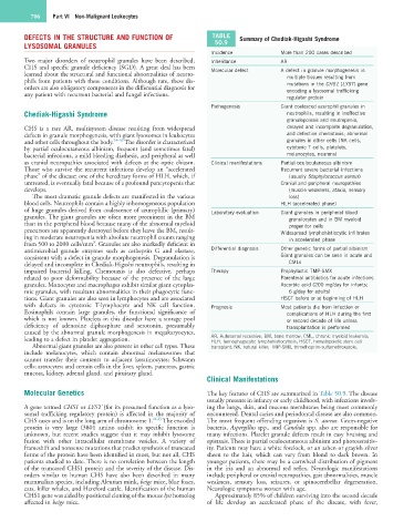
DEFECTS IN THE STRUCTURE AND FUNCTION OF TABLE Summary of Chediak-Higashi Syndrome
LYSOSOMAL GRANULES 50.9
Incidence More than 200 cases described
Two major disorders of neutrophil granules have been described, Inheritance AR
CHS and specific granule deficiency (SGD). A great deal has been Molecular defect A defect in granule morphogenesis in
learned about the structural and functional abnormalities of neutro- multiple tissues resulting from
phils from patients with these conditions. Although rare, these dis- mutations in the CHS1 (LYST) gene
orders are also obligatory components in the differential diagnosis for encoding a lysosomal trafficking
any patient with recurrent bacterial and fungal infections.
regulator protein
Pathogenesis Giant coalesced azurophil granules in
Chediak-Higashi Syndrome neutrophils, resulting in ineffective
granulopoiesis and neutropenia,
CHS is a rare AR, multisystem disease resulting from widespread delayed and incomplete degranulation,
defects in granule morphogenesis, with giant lysosomes in leukocytes and defective chemotaxis; abnormal
and other cells throughout the body. 24–26 The disorder is characterized granules in other cells (NK cells,
by partial oculocutaneous albinism, frequent (and sometimes fatal) cytotoxic T cells, platelets,
bacterial infections, a mild bleeding diathesis, and peripheral as well melanocytes, neurons)
as cranial neuropathies associated with defects at the optic chiasm. Clinical manifestations Partial oculocutaneous albinism
Those who survive the recurrent infections develop an “accelerated Recurrent severe bacterial infections
phase” of the disease; one of the hereditary forms of HLH, which, if (usually Staphylococcus aureus)
untreated, is eventually fatal because of a profound pancytopenia that Cranial and peripheral neuropathies
develops. (muscle weakness, ataxia, sensory
The most dramatic granule defects are manifested in the various loss)
blood cells. Neutrophils contain a highly inhomogeneous population HLH (accelerated phase)
of huge granules derived from coalescence of azurophilic (primary) Laboratory evaluation Giant granules in peripheral blood
granules. The giant granules are often more prominent in the BM granulocytes and in BM myeloid
than in the peripheral blood because many of the abnormal myeloid progenitor cells
precursors are apparently destroyed before they leave the BM, result- Widespread lymphohistiocytic infiltrates
ing in moderate neutropenia with absolute neutrophil counts ranging in accelerated phase
3
from 500 to 2000 cells/mm . Granules are also markedly deficient in
antimicrobial granule enzymes such as cathepsin G and elastase, Differential diagnosis Other genetic forms of partial albinism
consistent with a defect in granule morphogenesis. Degranulation is Giant granules can be seen in acute and
delayed and incomplete in Chediak-Higashi neutrophils, resulting in CMLs
impaired bacterial killing. Chemotaxis is also defective, perhaps Therapy Prophylactic TMP-SMX
related to poor deformability because of the presence of the large Parenteral antibiotics for acute infections
granules. Monocytes and macrophages exhibit similar giant cytoplas- Ascorbic acid (200 mg/day for infants;
mic granules, with resultant abnormalities in their phagocytic func- 6 g/day for adults)
tions. Giant granules are also seen in lymphocytes and are associated HSCT before or at beginning of HLH
with defects in cytotoxic T-lymphocyte and NK cell function. Prognosis Most patients die from infection or
Eosinophils contain large granules, the functional significance of complications of HLH during the first
which is not known. Platelets in this disorder have a storage pool or second decade of life unless
deficiency of adenosine diphosphate and serotonin, presumably transplantation is performed
caused by the abnormal granule morphogenesis in megakaryocytes,
leading to a defect in platelet aggregation. AR, Autosomal recessive; BM, bone marrow; CML, chronic myeloid leukemia;
HLH, hemophagocytic lymphohistiocytosis; HSCT, hematopoietic stem cell
Abnormal giant granules are also present in other cell types. These transplant; NK, natural killer; TMP-SMX, trimethoprim-sulfamethoxazole.
include melanocytes, which contain abnormal melanosomes that
cannot transfer their contents to adjacent keratinocytes; Schwann
cells; astrocytes; and certain cells in the liver, spleen, pancreas, gastric
mucosa, kidney, adrenal gland, and pituitary gland.
Clinical Manifestations
Molecular Genetics The key features of CHS are summarized in Table 50.9. The disease
usually presents in infancy or early childhood, with infections involv-
A gene termed CHS1 or LYST (for its presumed function as a lyso- ing the lungs, skin, and mucous membranes being most commonly
somal trafficking regulatory protein) is affected in the majority of encountered. Dental caries and periodontal disease are also common.
CHS cases and is on the long arm of chromosome 1. 24,25 The encoded The most frequent offending organism is S. aureus. Gram-negative
protein is very large (3801 amino acids); its specific function is bacteria, Aspergillus spp., and Candida spp. also are responsible for
unknown, but recent studies suggest that it may inhibit lysosome many infections. Platelet granule defects result in easy bruising and
fusion with other intracellular membrane vesicles. A variety of epistaxis. There is partial oculocutaneous albinism and photosensitiv-
frameshift and nonsense mutations that predict synthesis of truncated ity. Patients may have a white forelock, or an ashen or grayish silver
forms of the protein have been identified in most, but not all, CHS sheen to the hair, which can vary from blond to dark brown. In
patients studied to date. There is no correlation between the length younger patients, there may be a cartwheel distribution of pigment
of the truncated CHS1 protein and the severity of the disease. Dis- in the iris and an abnormal red reflex. Neurologic manifestations
orders similar to human CHS have also been described in many include peripheral or cranial neuropathies, gait abnormalities, muscle
mammalian species, including Aleutian mink, beige mice, blue foxes, weakness, sensory loss, seizures, or spinocerebellar degeneration.
cats, killer whales, and Hereford cattle. Identification of the human Neurologic symptoms worsen with age.
CHS1 gene was aided by positional cloning of the mouse lyst homolog Approximately 85% of children surviving into the second decade
affected in beige mice. of life develop an accelerated phase of the disease, with fever,