
Page 843 - Hematology_ Basic Principles and Practice ( PDFDrive )
P. 843
726 Part VI Non-Malignant Leukocytes
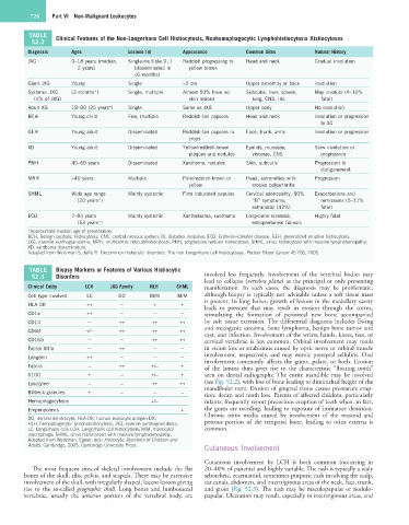
TABLE Clinical Features of the Non-Langerhans Cell Histiocytosis, Nonhemophagocytic Lymphohistiocytosis Histiocytoses
52.2
Diagnosis Ages Lesions (n) Appearance Common Sites Natural History
JXG 0–18 years (median, Single:multiple 9 : 1 Reddish progressing to Head and neck Gradual involution
2 years) (disseminated in yellow brown
<6 months)
Giant JXG Young Single >2 cm Upper extremity or back Involution
Systemic JXG (3 months*) Single, multiple Almost 50% have no Subcutis, liver, spleen, May involute (4–10%
(4% of JXG) skin lesions lung, CNS, iris fatal)
Adult XG 18–80 (35 years*) Single Same as JXG Upper body No involution
BCH Young child Few, multiple Reddish-tan papules Head and neck Involution or progression
to XG
GEH Young adult Disseminated Reddish-tan papules in Face, trunk, arms Involution or progression
crops
XD Young adult Disseminated Yellow/reddish-brown Eyelids, mucosae, Slow involution or
plaques and nodules viscerae, CNS progression
PNH 40–60 years Disseminated Xanthoma, nodules Skin, subcutis Progression to
disfigurement
MRH >40 years Multiple Pink/reddish-brown or Head, extremities with Progression
yellow erosive polyarthritis
SHML Wide age range Mainly systemic Firm indurated papules Cervical adenopathy, 80% Exacerbations and
(20 years*) “B” symptoms, remissions (5–11%
extranodal (43%) fatal)
ECD 7–84 years Mainly systemic Xanthelasma, xanthoma Long-bone sclerosis, Highly fatal
(53 years*) retroperitoneal fibrosis
*Approximate median age of presentation.
BCH, Benign cephalic histiocytosis; CNS, central nervous system; DI, diabetes insipidus; ECD, Erdheim–Chester disease; GEH, generalized eruptive histiocytosis;
JXG, juvenile xanthogranuloma; MRH, multicentric reticulohistiocytosis; PNH, progressive nodular histiocytosis; SHML, sinus histiocytosis with massive lymphadenopathy;
XD, xanthoma disseminatum.
Adapted from Weitzman S, Jaffe R: Uncommon histiocytic disorders: The non-Langerhans cell histiocytoses. Pediatr Blood Cancer 45:256, 2005.
TABLE Biopsy Markers or Features of Various Histiocytic
52.3 Disorders involved less frequently. Involvement of the vertebral bodies may
lead to collapse (vertebra plana) as the principal or only presenting
Clinical Entity LCH JXG Family HLH SHML manifestation. In such cases, the diagnosis may be problematic,
Cell type involved LC DD M/M M/M although biopsy is typically not advisable unless a soft tissue mass
HLA-DR ++ − + + is present. In long bones, growth of lesions in the medullary cavity
leads to pressure that may result in erosion through the cortex,
CD1a ++ − − − stimulating the formation of periosteal new bone accompanied
CD14 − ++ ++ ++ by soft tissue extension. The differential diagnosis includes Ewing
and osteogenic sarcoma, bone lymphoma, benign bone tumor and
CD68 +/− ++ ++ ++
cyst, and infection. Involvement of the wrists, hands, knees, feet, or
CD163 − − ++ ++ cervical vertebrae is less common. Orbital involvement may result
Factor XIIIa − ++ − − in vision loss or strabismus caused by optic nerve or orbital muscle
Langerin ++ − − − involvement, respectively, and may mimic preseptal cellulitis. Oral
involvement commonly affects the gums, palate, or both. Erosion
Fascin − ++ +/− + of the lamina dura gives rise to the characteristic “floating tooth”
S100 + − +/− + seen on dental radiographs. The entire mandible may be involved
Lysozyme − − ++ ++ (see Fig. 52.2), with loss of bone leading to diminished height of the
Birbeck granules + − − − mandibular rami. Erosion of gingival tissue causes premature erup-
tion, decay, and tooth loss. Parents of affected children, particularly
Hemophagocytosis +/− infants, frequently report precocious eruption of teeth when, in fact,
Emperipolesis + the gums are receding, leading to exposure of immature dentition.
Chronic otitis media caused by involvement of the mastoid and
DD, dermal dendrocyte; HLA-DR, human leukocyte antigen-DR;
HLH, hemophagocytic lymphohistiocytosis; JXG, juvenile xanthogranuloma; petrous portion of the temporal bone, leading to otitis externa is
LC, Langerhans cell; LCH, Langerhans cell histiocytosis; M/M, monocyte/ common.
macrophage; SHML, sinus histiocytosis with massive lymphadenopathy.
Adapted from Weitzman, Egeler, eds: Histiocytic Disorders of Children and
Adults. Cambridge, 2005, Cambridge University Press.
Cutaneous Involvement
Cutaneous involvement by LCH is both common (occurring in
The most frequent sites of skeletal involvement include the flat 20–40% of patients) and highly variable. The rash is typically a scaly
bones of the skull, ribs, pelvis, and scapula. There may be extensive seborrheic, eczematoid, sometimes purpuric rash involving the scalp,
involvement of the skull, with irregularly shaped, lucent lesions giving ear canals, abdomen, and intertriginous areas of the neck, face, trunk,
rise to the so-called geographic skull. Long bones and lumbosacral and groin (Fig. 52.3). The rash may be maculopapular or nodulo-
vertebrae, usually the anterior portion of the vertebral body, are papular. Ulceration may result, especially in intertriginous areas, and