
Page 844 - Hematology_ Basic Principles and Practice ( PDFDrive )
P. 844
Chapter 52 Histiocytic Disorders 727
P P
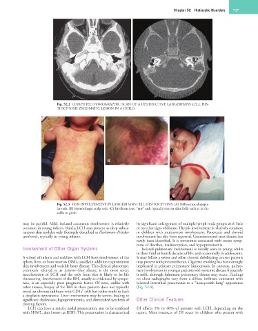
Fig. 52.2 COMPUTED TOMOGRAPHIC SCAN OF A DESTRUCTIVE LANGERHANS CELL HIS-
TIOCYTOSIS ZYGOMATIC LESION IN A CHILD.
B
A C
Fig. 52.3 SKIN INVOLVEMENT IN LANGERHANS CELL HISTIOCYTOSIS. (A) Diffuse maculopapu-
lar rash. (B) Hemorrhagic scalp rash. (C) Erythematous, “raw” rash typically seen in skin folds such as in the
axilla or groin.
may be painful. Mild, isolated cutaneous involvement is relatively by significant enlargement of multiple lymph node groups with little
common in young infants. Rarely, LCH may present as deep subcu- or no other signs of disease. Thymic involvement is relatively common
taneous skin nodules only (formerly described as Hashimoto–Pritzker in children with multisystem involvement. Pancreatic and thyroid
syndrome), typically in young infants. involvement has also been reported. Gastrointestinal tract disease has
rarely been identified. It is sometimes associated with severe symp-
toms of diarrhea, malabsorption, and hypoproteinemia.
Involvement of Other Organ Systems Isolated pulmonary involvement is usually seen in young adults
in their third or fourth decades of life, and occasionally in adolescents.
A subset of infants and toddlers with LCH have involvement of the It may follow a severe and often chronic debilitating course; patients
spleen, liver, or bone marrow (BM), usually in addition to prominent may present with pneumothorax. Cigarette smoking has been strongly
skin involvement and variable bony disease. This clinical phenotype, implicated in primary pulmonary histiocytosis. In contrast, pulmo-
previously referred to as Letterer–Siwe disease, is the most severe nary involvement in younger patients with systemic disease frequently
manifestation of LCH and the only form that is likely to be life is mild, although fulminant pulmonary disease may occur. Findings
threatening. Involvement of the BM, usually as evidenced by cytope- on chest radiographs vary from a diffuse infiltrate consistent with
nias, is an especially poor prognostic factor. Of note, unlike with bilateral interstitial pneumonia to a “honeycomb lung” appearance
other tissues, biopsy of the BM in these patients does not typically (Fig. 52.4).
+
reveal an obvious infiltrate with CD1a cells but rather tends to have
a dysplastic appearance. Liver involvement may be severe, leading to
significant cholestasis, hypoproteinemia, and diminished synthesis of Other Clinical Features
clotting factors.
LCH can have a strictly nodal presentation, not to be confused DI affects 5% to 40% of patients with LCH, depending on the
with SHML, also known as RDD. This presentation is characterized report. Most instances of DI occur in children who present with