
Page 865 - Hematology_ Basic Principles and Practice ( PDFDrive )
P. 865
748 Part VI Non-Malignant Leukocytes
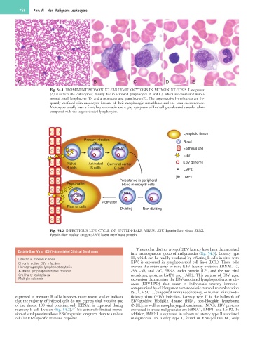
A B C D E
Fig. 54.1 PROMINENT MONONUCLEAR LYMPHOCYTOSIS IN MONONUCLEOSIS. Low power
(A) illustrates the leukocytosis, mainly due to activated lymphocytes (B and C), which are contrasted with a
normal small lymphocyte (D) and a monocyte and granulocyte (E). The large reactive lymphocytes are fre-
quently confused with monocytes because of their morphologic resemblance and the term mononucleosis.
Monocytes usually have a finer, lacy chromatin and a gray cytoplasm with small granules and vacuoles when
compared with the large activated lymphocytes.
Lymphoid tissue
Primary infection
B cell
EBNAs EBNA1 Epithelial cell
EBV
Naive Activated Germinal center EBV genome
B cells B cells B cells LMP2
LMP1
Reactivation Persistence in peripheral
blood memory B cells
EBNA1
Lytic genes
Activation
Plasma cells
Dividing Non-dividing
Fig. 54.2 INFECTIOUS LIFE CYCLE OF EPSTEIN-BARR VIRUS. EBV, Epstein-Barr virus; EBNA,
Epstein-Barr nuclear antigen; LMP, latent membrane protein.
Three other distinct types of EBV latency have been characterized
Epstein-Barr Virus (EBV)–Associated Clinical Syndromes
in a heterogeneous group of malignancies (Fig. 54.3). Latency type
Infectious mononucleosis III, which can be readily produced by infecting B cells in vitro with
Chronic active EBV infection EBV, is expressed in lymphoblastoid cell lines (LCL). These cells
Hemophagocytic lymphohistiocytosis express the entire array of nine EBV latency proteins: EBNA1, -2,
X-linked lymphoproliferative disease -3A, -3B, and -3C, EBNA leader protein (LP), and the two viral
Oral hairy leukoplakia membrane proteins LMP1 and LMP2. This pattern of EBV gene
Multiple sclerosis expression characterizes the EBV-associated lymphoproliferative dis-
eases (EBV-LPD) that occur in individuals severely immuno-
compromised by solid organ or hematopoietic stem cell transplantation
(SOT, HSCT), congenital immunodeficiency, or human immunode-
expressed in memory B cells; however, more recent studies indicate ficiency virus (HIV) infection. Latency type II is the hallmark of
that the majority of infected cells do not express viral proteins and EBV-positive Hodgkin disease (HD), non-Hodgkin lymphoma
of the almost 100 viral proteins, only EBNA1 is expressed during (NHL), as well as nasopharyngeal carcinoma (NPC). EBV proteins
2
memory B-cell division (Fig. 54.2). This extremely limited expres- expressed in these malignancies are EBNA1, LMP1, and LMP2. In
sion of viral proteins allows EBV to persist long term despite a robust addition, BARF1 is expressed in subsets of latency type II associated
cellular EBV-specific immune response. malignancies. In latency type I, found in EBV-positive BL, only