
Page 869 - Hematology_ Basic Principles and Practice ( PDFDrive )
P. 869
752 Part VI Non-Malignant Leukocytes
the majority of patients recover within 1 to 2 months. Patients usually Other Organ Involvement
have a positive direct Coombs test. Most common anti-I antibodies
are present; however, anti-I, anti-N, and Donath-Landsteiner anti- Although symptomatic heart disease with IM is uncommon, in one
bodies have also been reported. In addition to hemolysis, IM-associated cohort of patients, unspecific ST- and T-wave abnormalities were
anemia can be caused by erythroblastopenia. found in 6% of patients. Renal involvement manifested as micro-
scopic hematuria, and proteinuria is seen in 10% to 15% of patients;
Neutropenia however, significant renal dysfunction is rare. Airway compromise
Mild, self-limiting neutropenia is a common finding during the first due to hypertrophy of the adenoids and tonsils or mucosal inflam-
4 weeks of the disease. However, severe neutropenia associated with mation and edema is uncommon but potentially fatal.
fatal bacterial infections has been reported.
Thrombocytopenia Diagnosis
3
Mild thrombocytopenia (50,000 to 150,000/mm ) is a common
finding in patients with IM. It usually occurs within the first 2 Atypical lymphocytosis is the cardinal hematologic finding in IM
weeks of presentation and resolves within 2 months. Severe (Fig. 54.4). It develops during the first week of the illness and peaks
thrombocytopenia with overt bleeding is rare; however, death from between the second and third week. Atypical lymphocytes represent
intracranial hemorrhage has been described. The etiology of the 60% to 70% of the total white cell count, which ranges between
3
3
thrombocytopenia is not completely understood, and a variety of 12,000/mm and 18,000/mm . In general, the atypical lymphocytes
explanations have been suggested. Because bone marrow examination are large and vary in size. Nuclei are large and eccentrically placed;
shows normal or increased numbers of megakaryocytes, peripheral the cytoplasm is basophilic, and vacuoles are often present. The
platelet destruction is most likely due to the presence of antiplatelet variable morphologic pattern of atypical lymphocytes in IM distin-
antibodies or platelet pooling and destruction within an enlarged guishes them from the monotonous appearance of immature leukemic
spleen. blasts. Atypical lymphocytosis is not pathognomonic for IM and is
associated with other diseases such as acute viral hepatitis, CMV
infections, mumps, toxoplasmosis, rubella, roseola, and drug
Splenic Rupture reactions.
The diagnosis of EBV infection depends on serologic testing. Tests
Splenic rupture occurs predominately in males, with an incidence of for heterophile antibodies, including the monospot test and slide
1/1000 to 1/3000 (Fig. 54.5). The incidence of rupture is highest agglutination tests, are routinely available. The results of these tests
in the second and third week of illness and can be the first sign of are often negative in children less than 4 years of age, but they identify
IM. Clinical symptoms include abdominal pain or pain referred to 90% of cases in older children and adults. Of the available EBV-
either shoulder. Because abdominal pain is an unusual symptom of specific serologic tests, VCA-IgM antibodies are most commonly
uncomplicated IM, a splenic rupture should be strongly considered determined to diagnose primary EBV infection in heterophile-
in IM cases when abdominal pain is reported. Although it is a life- negative IM cases; determining antibodies against EA may also be
threatening complication, with current management the mortality helpful (Table 54.1). VCA-IgG antibodies are positive during acute
rate is very low. infections as well as the convalescent period. The presence of anti-
EBNA antibodies excludes an acute infection. Isolation of EBV from
throat washings is feasible; however, it is of little diagnostic value
Neurologic Complications because 10% to 20% of healthy adult EBV carriers may shed the
virus.
Neurologic complications develop usually during the first 2 weeks
of IM and may be the only manifestation of IM. EBV infec-
tion can cause a wide spectrum of neurologic diseases, including Differential Diagnosis
encephalitis, meningitis, Guillain-Barré syndrome, acute transverse
myelitis, and peripheral neuritis. Patients with neurologic compli- In the majority of cases, the diagnosis of IM is straightforward. The
cations have an excellent outcome, with most patients recovering differential diagnosis includes streptococcal and nonstreptococcal
completely. pharyngitis, acute infections with CMV, human herpesvirus 6,
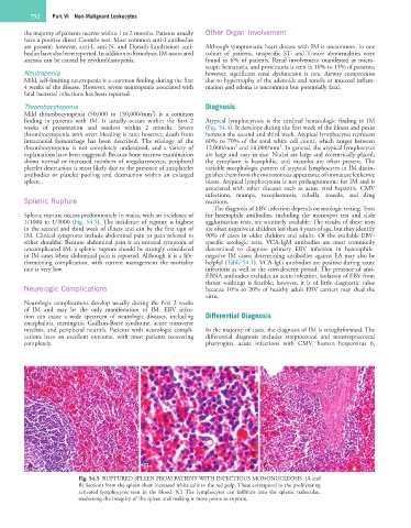
A B C
Fig. 54.5 RUPTURED SPLEEN FROM PATIENT WITH INFECTIOUS MONONUCLEOSIS. (A and
B) Sections from the spleen show increased white cells in the red pulp. These correspond to the proliferating
activated lymphocytes seen in the blood. (C) The lymphocytes can infiltrate into the splenic trabeculae,
weakening the integrity of the spleen and making it more prone to rupture.