
Page 866 - Hematology_ Basic Principles and Practice ( PDFDrive )
P. 866
Chapter 54 Infectious Mononucleosis and Other Epstein-Barr Virus–Associated Diseases 749
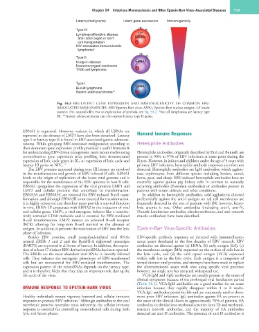
Latency/malignancy Latent gene expression Immunogenicity
Type III
Lymphoproliferative disease LP
after solid organ or stem EBNAs
cell transportation
HIV-associated immunoblastic
lymphoma ∗
Type II
Hodgkin disease EBNA1
Nasopharyngeal carcinoma
T/NK-cell lymphoma
Type I
Burkitt lymphoma EBNA1
Gastric adenocarcinoma ∗∗
Fig. 54.3 EBV-LATENT GENE EXPRESSION AND IMMUNOGENICITY OF COMMON EBV-
ASSOCIATED MALIGNANCIES. EBV, Epstein-Barr virus; EBNA, Epstein-Barr nuclear antigen; LP, leader
protein; NK, natural killer. For an explanation of symbols, see Fig. 54.2. *Not all lymphomas are latency type
III. **Gastric adenocarcinoma can also express latency type II genes.
EBNA1 is expressed. However, variants in which all EBNAs are
expressed in the absence of LMP1 have also been described. Latency Humoral Immune Responses
type I or latency type II is found in EBV-associated gastric adenocar-
cinoma. While grouping EBV-associated malignancies according to Heterophile Antibodies
their dominant gene expression profile provided a useful framework
for understanding EBV-driven oncogenesis, more recent studies using Heterophile antibodies, originally described by Paul and Bunnell, are
comprehensive gene expression array profiling have demonstrated present in 90% to 95% of EBV infections at some point during the
expression of lytic cycle genes in BL, or expression of lytic cycle and illness. However, in infants and children under the age of 4 years with
latency III genes in NPC. 5,6 primary EBV infection, heterophile antibody responses are often not
The EBV proteins expressed during type III latency are involved detected. Heterophile antibodies are IgM antibodies, which aggluti-
in the transformation and growth of EBV-infected B cells. EBNA1 nate erythrocytes from different species including bovine, camel,
binds to the origin of replication of the latent viral genome and is horse, goat, and sheep. EBV-induced heterophile antibodies have no
responsible for the maintenance of the EBV episome in host B cells. reactivity against guinea pig kidney cells in contrast to naturally
EBNA2 upregulates the expression of the viral proteins LMP1 and occurring antibodies (Forssman antibodies) or antibodies present in
LMP2 and cellular proteins that contribute to transformation. patients with serum sickness and other conditions.
EBNA3A and EBNA3C are essential for EBV-induced B-cell trans- In addition to heterophile antibodies, cold agglutinins directed
formation, and although EBNA3B is not essential for transformation, preferentially against the anti-I antigen on red cell membranes are
it is highly conserved and therefore must provide a survival function frequently detected in the sera of patients with IM; however, hemo-
in vivo. EBNA-LP cooperates with EBNA2 in the induction of viral lytic anemia is rare. Other antibodies (including anti-I, anti-N,
and cellular genes. LMP1, a viral oncogene, behaves like a constitu- Donath-Landsteiner antibodies, platelet antibodies, and anti–smooth
tively activated CD40 molecule and is essential for EBV-mediated muscle antibodies) have been described.
B-cell transformation. LMP2 mimics an activated B-cell receptor
(BCR) allowing for long-term B-cell survival in the absence of
antigen. In addition, it prevents the reactivation of EBV into the lytic Epstein-Barr Virus-Specific Antibodies
phase of infection.
Besides EBV proteins, small nonpolyadenylated viral RNAs EBV-specific antibody responses are detected with immunofluores-
termed EBERs 1 and 2 and the BamHI-A rightward transcripts cence assays developed in the first decades of EBV research. EBV
(BARTS) are expressed in all forms of latency. In addition, the expres- antibodies are directed against (a) EBNA, (b) early antigen (EA), (c)
sion of at least 17 distinct EBV-derived microRNAs has been reported. the membrane antigen (MA) expressed on the surface of cells late in
The EBERs are the most abundant viral RNAs in latently infected the lytic cycle, and (d) the viral capsid antigen (VCA) expressed
cells. They enhance the oncogenic phenotype of EBV-transformed within cells late in the lytic cycle. Each antigen is a composite of
cells but are nonessential for EBV-mediated transformation. The several distinct viral proteins, and attempts have been made to replace
expression pattern of the microRNAs depends on the latency type, the aforementioned assays with tests using specific viral proteins;
and it is therefore likely that they play an important role during the however, no single test has attracted widespread use.
life cycle of the virus. VCA-IgM and -IgG antibodies are usually present at the onset of
clinical symptoms because of the prolonged viral incubation period
(Table 54.1). VCA-IgM antibodies are a good marker for an acute
IMMUNE RESPONSE TO EPSTEIN-BARR VIRUS infection because they rapidly disappear within 4 to 8 weeks.
VCA-IgG antibodies persist for life and are commonly used to docu-
Healthy individuals mount vigorous humoral and cellular immune ment prior EBV infection. IgG antibodies against EA are present at
7
responses to primary EBV infection. Although antibodies to the viral the onset of the clinical illness in approximately 70% of patients. EA
membrane proteins neutralize virus infectivity, the cellular immune antibodies are divided into methanol-sensitive (anti-D) and methanol-
response is essential for controlling virus-infected cells during both resistant (anti-R) antibodies, and the majority of EA antibodies
lytic and latent phases. detected are anti-D antibodies. The presence of anti-D antibodies is