
Page 868 - Hematology_ Basic Principles and Practice ( PDFDrive )
P. 868
Chapter 54 Infectious Mononucleosis and Other Epstein-Barr Virus–Associated Diseases 751
administration was safe, no data are available if the vaccine boosted Complications of Primary Epstein-Barr Virus Infections
LMP2-specific T-cell responses.
The incidence of complications associated with primary EBV infec-
tion is low, although any organ system can be affected.
INFECTIOUS MONONUCLEOSIS
Epidemiology Hematologic Complications
EBV infections occur worldwide and in most populations 90% to Patients with IM may present with a wide range of hematologic
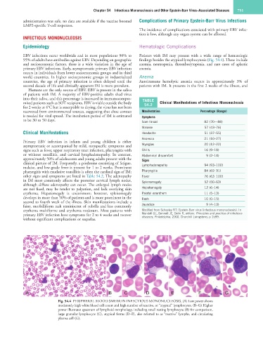
95% of adults have antibodies against EBV. Depending on geographic findings besides the atypical lymphocytosis (Fig. 54.4). These include
and socioeconomic factors, there is a wide variation in the age of anemia, neutropenia, thrombocytopenia, and rare cases of aplastic
primary EBV infections. Early, asymptomatic primary EBV infection anemia.
occurs in individuals from lower socioeconomic groups and in third
world countries. In higher socioeconomic groups in industrialized Anemia
countries, the age of primary infection is often delayed until the Autoimmune hemolytic anemia occurs in approximately 3% of
second decade of life and clinically apparent IM is more prevalent. patients with IM. It presents in the first 2 weeks of the illness, and
Humans are the only source of EBV. EBV is present in the saliva
of patients with IM. A majority of EBV-positive adults shed virus
into their saliva, and this percentage is increased in immunocompro- TABLE
mised patients such as SOT recipients. EBV is viable outside the body 54.2 Clinical Manifestations of Infectious Mononucleosis
for 2 weeks at 4°C but is susceptible to drying; the virus has not been
recovered from environmental sources, suggesting that close contact Manifestation Percentage (Range)
is needed for viral spread. The incubation period of IM is estimated Symptoms
to be 30 to 50 days. Sore throat 82 (70––88)
Malaise 57 (43–76)
Clinical Manifestations Headache 51 (37–55)
Anorexia 21 (10–27)
Primary EBV infection in infants and young children is either
asymptomatic or accompanied by mild, nonspecific symptoms and Myalgias 20 (12–22)
signs such as fever, upper respiratory tract infection, pharyngitis with Chills 16 (9–18)
or without tonsillitis, and cervical lymphadenopathy. In contrast, Abdominal discomfort 9 (2–14)
approximately 50% of adolescents and young adults present with the Signs
clinical picture of IM. Frequently, a prodrome consisting of fatigue, Lymphadenopathy 94 (93–100)
malaise, and low-grade fever is present for 1 to 2 weeks. Prominent
pharyngitis with exudative tonsillitis is often the cardinal sign of IM; Pharyngitis 84 (69–91)
other signs and symptoms are listed in Table 54.2. The adenopathy Fever 76 (63–100)
in IM most commonly affects the posterior cervical lymph nodes, Splenomegaly 52 (50–63)
although diffuse adenopathy can occur. The enlarged lymph nodes
are not fixed, may be tender to palpation, and lack overlying skin Hepatomegaly 12 (6–14)
erythema. Hepatomegaly is uncommon; however, splenomegaly Palatal enanthem 11 (5–13)
develops in more than 50% of patients and is more prominent in the Rash 10 (0–15)
second to fourth week of the illness. Skin manifestations include a
faint, morbilliform rash reminiscent of rubella and less commonly Jaundice 9 (4–10)
erythema multiforme and erythema nodosum. Most patients with Modified from Schooley RT: Epstein-Barr virus (infectious mononucleosis). In
primary EBV infection have symptoms for 2 to 4 weeks and recover Mandell GL, Bennett JE, Dolin R, editors: Principles and practice of infectious
diseases, Philadelphia, 2000, Churchill Livingstone, p 1599.
without significant complications or sequelae.
A B C D E F G
Fig. 54.4 PERIPHERAL BLOOD SMEAR IN INFECTIOUS MONONUCLEOSIS. (A) Low power shows
moderately high white blood cell count and high number of reactive, or “atypical” lymphocytes. (B–G) Higher
power illustrates spectrum of lymphoid morphology, including small resting lymphocyte (B) for comparison,
large granular lymphocyte (C), atypical forms (D–F), also referred to as “reactive” lymphs, and circulating
plasma cell (G).