
Page 886 - Hematology_ Basic Principles and Practice ( PDFDrive )
P. 886
Chapter 55 Progress in the Classification of Hematopoietic and Lymphoid Neoplasms 769
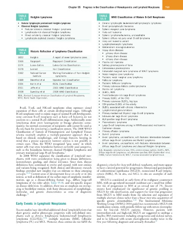
TABLE Hodgkin Lymphoma TABLE WHO Classification of Mature B-Cell Neoplasms
55.7 55.9
• Nodular lymphocyte predominant Hodgkin lymphoma • Chronic lymphocytic leukemia/small lymphocytic lymphoma
• Classical Hodgkin lymphoma • B-cell prolymphocytic leukemia
• Nodular sclerosis classical Hodgkin lymphoma • Splenic marginal zone lymphoma
• Lymphocyte-rich classical Hodgkin lymphoma • Hairy cell leukemia
• Mixed cellularity classical Hodgkin lymphoma • Splenic lymphoma/leukemia, unclassifiable
• Lymphocyte-depleted classical Hodgkin lymphoma • Splenic diffuse red pulp small B-cell lymphoma
• Hairy cell leukemia variant
• Lymphoplasmacytic lymphoma
• Waldenström macroglobulinemia
TABLE Historic Reflection of Lymphoma Classification • Heavy chain diseases
55.8 • µHeavy chain disease
• γHeavy chain disease
1832 Hodgkin A report of seven lymphoma cases
• αHeavy chain disease
1966 Rappaport Rappaport Classification • Plasma cell myeloma
1974 Lukes–Collins Lukes–Collins Classification • Solitary plasmacytoma of bone
1978 Lennert Keil Classification • Extraosseous plasmacytoma
• Extranodal marginal zone lymphoma of MALT lymphoma
1982 National Cancer Working Formulation of Non-Hodgkin • Nodal marginal zone lymphoma
Institute Lymphoma
• Pediatric nodal marginal zone lymphoma
1988 Stansfeld et al Updated Keil Classification • Follicular lymphoma
1994 Harris et al REAL Classification • Pediatric follicular lymphoma
• Primary cutaneous follicle centre lymphoma
2001 Jaffe et al 2001 WHO Classification
• Mantle cell lymphoma
2008 Swerdlow et al 2008 WHO Classification • DLBCL, NOS
REAL, Revised European-American Classification of Lymphoid Neoplasms; • T-cell/histiocyte–rich large B-cell lymphoma
WHO, World Health Organization. • Primary DLBCL of the CNS
• Primary cutaneous DLBCL, leg type
• EBV-positive DLBCL of the elderly
B-cell, T-cell, and NK-cell neoplasms often represent clonal • DLBCL associated with chronic inflammation
expansion of these cells at certain developmental stages. Although • Lymphomatoid granulomatosis
B-cell neoplasms tend to mimic stages of normal B-cell development, • Primary mediastinal (thymic) large B-cell lymphoma
some common B-cell neoplasms such as hairy cell leukemia do not • Intravascular large B-cell lymphoma
conform to a normal B-cell differentiation stage. Additionally, some • ALK-positive large B-cell lymphoma
lymphomas show overt heterogeneity or lineage plasticity; conse- • Plasmablastic lymphoma
quently, the normal counterpart of neoplastic cells cannot be used as • Large B-cell lymphoma arising in HHV8-associated multicentric
the sole basis for developing a classification system. The 2008 WHO Castleman disease
Classification of Tumors of Hematopoietic and Lymphoid Tissues • Primary effusion lymphoma
schema routinely employs a multiple-parameter approach that is • Burkitt lymphoma
based on clinical, morphologic, and biologic features, keeping in • B-cell lymphoma, unclassifiable, with features intermediate between
mind that a precise separation between entities is not possible in diffuse large B-cell lymphoma and Burkitt lymphoma
certain cases. Thus, the WHO recognized “gray zones” in which • B-cell lymphoma, unclassifiable, with features intermediate between
tumor cells may cross boundaries between currently used categories, diffuse large B-cell lymphoma and classical Hodgkin lymphoma
such as the boundaries between classical Hodgkin lymphoma and ALK, Anaplastic lymphoma kinase; CNS, central nervous system; DLBCL, NOS,
primary mediastinal large B-cell lymphoma. 4 Diffuse large B-cell lymphoma, not otherwise specified; EBV, Epstein-Barr virus;
In 2008 WHO expanded the classification of lymphoid neo- HHV8, human herpesvirus-8; MALT, mucosa-associated lymphoid tissue.
plasms, with more consideration being given to disease definitions,
nomenclature, grading, and clinical relevance. Since then, disease
definitions have continued to evolve and expand, with new entities diagnostic criteria for these well-defined neoplasms, and many appear
and variants being recognized. Both clinical and laboratory research to have a limited potential for progression. Monoclonal gammopathy
findings provided new insights that are relevant to these emerging of undetermined significance (MGUS), monoclonal B-cell lympho-
concepts. 23,24 Current areas of development focus on early or in situ cytosis (MBL), FL in situ, and MCL in situ are examples of such
lesions, as well as definition of the earlier steps of neoplastic transfor- entities.
mation, age as a disease-defining feature (e.g., diffuse large-cell MGUS is considered an early form of its malignant counterpart
lymphoma of the older adult; Table 55.9), and site-specific impact MM, with an age-related increased incidence and a small but defini-
on disease definition. In addition, there was an emphasis on overlap- tive risk of progression to MM at an annual rate of 1%. Recent
ping or borderline entities, with fuzzy demarcation of morphologic, reports have emphasized the significance of genetic profiling in
molecular, and genetic characteristics as areas of diagnostic MGUS for risk stratification, and support the view that progression
challenge. 23 from MGUS to MM results from the selection and expansion of
multiple aberrant clones rather than a linear step-wise acquisition of
specific genetic abnormalities. 27,28 The International Myeloma
Early Events in Lymphoid Neoplasms Working Group (IMWG) 2010 guidelines recommend a MGUS risk
stratification system, with periodic follow-up with serum electropho-
Recent studies have identified additional clonal lymphoid lesions that resis for low-risk MGUS patients. However, patients with
share genetic and/or phenotypic properties with well-defined neo- intermediate-risk and high-risk MGUS are suggested to undergo a
plasms such as chronic lymphocytic leukemia/small lymphocytic baseline BM examination including cytogenetics and skeletal survey,
lymphoma (CLL/SLL), 25,26 multiple myeloma (MM), follicular and to be followed with serum electrophoresis studies twice in the
lymphoma (FL) and MCL. However, these entities do not fulfill the first year following diagnosis and annually thereafter. 29