
Page 888 - Hematology_ Basic Principles and Practice ( PDFDrive )
P. 888
Chapter 55 Progress in the Classification of Hematopoietic and Lymphoid Neoplasms 771
overexpression. Patients with dual overexpression of MYC and BCL2 Peripheral T-Cell Lymphoma
have a significantly poorer outcome compared with patients who
express only one or neither protein. In addition, concurrent MYC Peripheral T-cell lymphomas (PTCLs) encompass numerous entities
and BCL2 translocation, known as double-hit lymphoma, indicates a (Table 55.10) that are characterized by a poor prognosis, with the
subgroup of patients who are refractory to treatment and have a exception of histologic subtype “ALK-positive anaplastic large-cell
median survival of approximately 8 months. lymphoma.” Most PTCLs lack distinct genetic or biologic features,
The 2008 WHO classification emphasizes the importance of and the mechanisms underlying the pathogenesis of these lymphomas
integrating morphologic, immunophenotypic, and molecular data to are not yet fully understood. However, development of genomic
make a final diagnosis. This integration has refined our ability to high-throughput profiling techniques now allows us to extensively
diagnose several entities such as DLBCL and Burkitt lymphoma identify the molecular abnormalities present in these entities. The
(BL). The 2008 WHO classification eliminated the variant category diagnosis of many cases is challenging even for expert hematopatholo-
of atypical BL, which had been included in the 2001 WHO classifica- gists, and more than a third of the cases cannot be further classified
+
+
tion. Thus, a case with the typical BL phenotype (CD20 , CD10 , and consequently are relegated to a “waste basket” category PTCL-
−
BCL2 ) and genotype (so-called MYC-simple or MYC/IG in the NOS. Other frequently encountered entities are angioimmunoblastic
absence of other major cytogenetic anomalies) may be classified as T-cell lymphoma (AITL), and anaplastic large cell lymphoma
4
BL even if there is some variability in the morphology of the neo- (ALCL). AITL and approximately 20% of PTCL-NOS show phe-
plastic cells. In addition, the 2008 WHO classification recognizes a notypic features of T-cell follicular helper (Tfh) cells; and share a
group of high-grade B-cell lymphomas that are not readily classified spectrum of genetic abnormalities such as TET2 and DNMT3A, as
as either BL or DLBCL. This provisional category is termed B-cell well as mutations in the motility and adhesion gene RHOA. 41
+
lymphoma, unclassifiable, with features that are intermediate between Despite morphologic and phenotypic similarities ALK ALCL is
4
DLBCL and BL, including double-hit lymphomas. In the 2016 considered a distinct entity that must be distinguished from the entity
−
WHO classification DLBCL with MYC and BCL2 and/or BCL6 of ALK ALCL given the clinical and biologic differences. In the 2016
−
rearrangements will be included in a single category to be designated update to the WHO classification, ALK ALCL are no longer provi-
high grade B cell lymphoma (HGBL), with MYC and BCL2 and/or sional entities and strict criteria are required for the diagnosis of
−
BCL6 rearrangements. The category of BCLU will be eliminated. ALK ALCL because CD30 may be expressed in a variety of PTCL
Cases that appear blastoid or cases intermediate between DLBCL and subtypes.
BL, but which lack a MYC and BCL2 and/or BCL6 rearrangement, Three variants of primary cutaneous PTCL were introduced in
will be placed in the category of HGBL, NOS. The 2008 WHO the 2008 WHO classification; primary cutaneous gamma-delta T-cell
+
classification recognizes another provisional category of B-cell neo- lymphoma, primary cutaneous CD4 small/medium T-cell lymphoma
plasms with features that are intermediate between DLBCL and as a provisional entity, and primary cutaneous aggressive epidermo-
+
classical Hodgkin lymphoma (CHL). These tumors occur predomi- tropic CD8 cytotoxic T-cell lymphoma. Cutaneous gamma-delta
nantly in young men and appear to be more aggressive than either T-cell lymphomas have a diverse histologic and clinical spectrum and
primary mediastinal large B-cell lymphoma or nodular sclerosis CHL. may display a panniculitis-like pattern. However, this disease has a
There are other settings in which the distinction between DLBCL much poorer prognosis than subcutaneous panniculitis-like T-cell
and CHL is challenging. For example, some EBV-associated B-cell lymphoma, which is defined as a lymphoma exclusively of alpha-beta
lymphomas may exhibit features that closely resemble or mimic phenotype in the 2008 WHO classification. Primary cutaneous
+
CHL. The borderline category should be used sparingly but is small/medium CD4 T-cell lymphoma is another lymphoma with
appropriate when a distinction between CHL and DLBCL is not
possible. 4
Several aggressive B-cell lymphomas have a distinct immunopro-
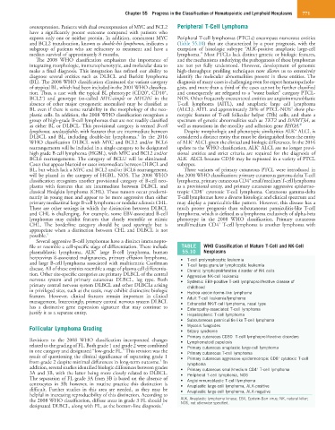
file or resemble a cell-specific stage of differentiation. These include TABLE WHO Classification of Mature T-Cell and NK-Cell
+
plasmablastic lymphoma, ALK large B-cell lymphoma, human 55.10 Neoplasms
herpesvirus 8-associated malignancies, primary effusion lymphoma, • T-cell prolymphocytic leukemia
and large B-cell lymphoma associated with multicentric Castleman • T-cell large granular lymphocytic leukemia
disease. All of these entities resemble a stage of plasma cell differentia- • Chronic lymphoproliferative disorder of NK cells
tion. Other site-specific categories are primary DLBCL of the central • Aggressive NK-cell leukemia
nervous system and primary cutaneous DLBCL, leg type. Both • Systemic EBV-positive T-cell lymphoproliferative disease of
primary central nervous system DLBCL and other DLBCLs arising childhood
in privileged sites, such as the testis, may exhibit distinctive biologic • Hydroa vacciniforme-like lymphoma
features. However, clinical features remain important in clinical • Adult T-cell leukemia/lymphoma
management. Interestingly, primary central nervous system DLBCL • Extranodal NK/T-cell lymphoma, nasal type
has a distinctive gene expression signature that may continue to • Enteropathy-associated T-cell lymphoma
justify it as a separate entity. • Hepatosplenic T-cell lymphoma
• Subcutaneous panniculitis-like T-cell lymphoma
Follicular Lymphoma Grading • Mycosis fungoides
• Sézary syndrome
• Primary cutaneous CD30 T-cell lymphoproliferative disorders
−
Revisions to the 2008 WHO classification incorporated changes • Lymphomatoid papulosis
related to the grading of FL. Both grade 1 and grade 2 were combined • Primary cutaneous anaplastic large-cell lymphoma
in one category and designated “low-grade FL.” This revision was the • Primary cutaneous T-cell lymphoma
result of questioning the clinical significance of separating grade 1 • Primary cutaneous aggressive epidermotropic CD8 cytotoxic T-cell
+
4
from grade 2 despite minimal differences in long-term outcome. In lymphoma
addition, several studies identified biologic differences between grades • Primary cutaneous small/medium CD4 T-cell lymphoma
+
3A and 3B, with the latter being more closely related to DLBCL. • Peripheral T-cell lymphoma, NOS
The separation of FL grade 3A from 3B is based on the absence of • Angioimmunoblastic T-cell lymphoma
centrocytes in 3B; however, in routine practice this distinction is • Anaplastic large-cell lymphoma, ALK-positive
difficult. Further studies in this area are needed, as they may be • Anaplastic large-cell lymphoma, ALK-negative
helpful in increasing reproducibility of this distinction. According to
the 2008 WHO classification, diffuse areas in grade 3 FL should be ALK, Anaplastic lymphoma kinase; EBV, Epstein-Barr virus; NK, natural killer;
NOS, not otherwise specified.
designated DLBCL, along with FL, as the bottom-line diagnosis. 4