
Page 889 - Hematology_ Basic Principles and Practice ( PDFDrive )
P. 889
772 Part VII Hematologic Malignancies
Tfh cell origin that presents commonly as an isolated lesion in the TABLE WHO Classification of Histiocytic and Dendritic Cell
42
head and neck region. In the 2016 WHO classification a change in 55.11 Neoplasms
the terminology of this entity is proposed—to primary cutaneous
+
small/medium CD4 T-cell lymphoproliferative disease instead of • Histiocytic sarcoma
lymphoma. • Langerhans cell histiocytosis
The 2008 WHO classification acknowledged that a variety of • Langerhans cell sarcoma
PTCLs can present with intestinal disease and that not all of these • Interdigitating dendritic cell sarcoma
cases are associated with celiac disease. Intestinal involvement can be • Follicular dendritic cell sarcoma
seen at presentation, and/or with progression, in extranodal NK/T- • Fibroblastic reticular cell tumor
cell lymphoma as well as in some gamma-delta T-cell lymphomas. • Intermediate dendritic cell tumor
The WHO classification required more stringent criteria to establish • Disseminated juvenile xanthogranuloma
a diagnosis known as enteropathy-associated T-cell lymphoma Specifi-
cally, in order to make the diagnosis of enteropathy-associated T-cell
lymphoma, evidence of celiac disease was required either at the
genetic level, with the appropriate HLA phenotype, or histologically, uncertain because many have been recognized only recently. Several
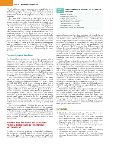
in the adjacent uninvolved small bowel mucosa. A new variant, entities are recognized in the 2008 WHO classification of histiocytic
termed the monomorphic variant of enteropathy-associated T-cell lym- and dendritic cell neoplasms (Table 55.11). Traditionally, these
phoma, or type II, was introduced into the 2008 WHO classification. tumors are placed into two basic categories based on their derivation
Cases exhibiting this variant have some distinctive immunopheno- from either BM precursors or mesenchymal cells. Histiocytic sarcoma
+
+
typic and genotypic features. The tumor cells are CD8 and CD56 , (HS), Langerhans cell histiocytosis (LCH), and interdigitating den-
and MYC amplifications are present in a subset of cases. The mono- dritic cell sarcoma (IDCS) are derived from BM precursors; while
morphic variant occurs in the setting of celiac disease but also occurs follicular dendritic cell sarcoma (FDCS), indeterminate dendritic cell
sporadically. 4 sarcoma, fibroblastic reticular cell tumors, and disseminated juvenile
xanthogranuloma are derived from stromal-derived dendritic cells or
are mesenchymal in origin. Although divergent differentiation from
Precursor Lymphoid Neoplasms marrow precursors is the normal histogenesis, hybrid and trans dif-
ferentiation from lymphoid clones has been proposed in some
The lymphoblastic neoplasms are derived from precursor cells or entities. 39
blasts, most of which are precursor B and T-cell neoplasms that Excisional biopsy is the preferred specimen choice from which to
present as leukemia. However, the designation of lymphoblastic render the diagnosis of these disorders. Consultation with an experi-
lymphoma is used when the neoplasm is confined to a mass lesion enced hematopathologist is often required, as morphologic review
without or with only minimal blood or BM involvement. The WHO and an adequate battery of immunohistochemical stains are the most
classification retains the convention that precursor neoplasms are important elements in making an accurate diagnosis of these entities
designated leukemia/lymphoma. When distinction between leukemia and in differentiating them from other, often-mistaken categories,
and lymphoma is required for clinical protocol eligibility in presenta- most commonly NHLs. The rarity of these disorders is the major
tion with a mass lesion and increased blasts in the BM, a threshold factor that makes this group of diseases difficult to accurately diagnose
of 25% blasts is used as the defining feature of leukemia. 4 and challenging to treat. Advances of immunohistochemistry have
The 2008 classification recognizes genetic features in the defini- contributed to an enhanced understanding of the biology of dendritic
tion of some forms of B-lymphoblastic leukemia (B-ALL). One such and histiocytic neoplasms, and have improved our ability to classify
+
example is Philadelphia chromosome-positive (Ph ) B-ALL, associ- and diagnose these disorders. For example, in contrast to LCH, IDCS
ated with BCR-ABL1, which is more common in adults than in are usually positive for S100 but negative for CD1a and langerin
children and is considered very high risk, regardless of other factors. (CD207). Unlike FDCS, IDCS do not express follicular dendritic
Deletions and other alterations in the IKZF1 (Ikaros) gene are adverse cell markers such as CD21 or CD35. 4
prognostic indicators in both Ph+ and Ph− patients with B-ALL. These entities can involve various organs, although most occur in
Another variant with distinctive clinical features at presentation is the lymph nodes and skin, with a unifocal or solitary presentation,
B-ALL/lymphoma with t(5;15)(q31;q32) (IL3-IGH). These patients and are associated with a good prognosis with surgical resection. On
present with a marked increase in eosinophils, which may mask a the other hand, cases with disseminated disease have shown a poor
relatively small number of blasts in the BM—a diagnostic pitfall outcome, although data on treatment options are limited. Nonethe-
worthy of note. The ongoing and increasing complexity highlights less, chemotherapy and referral to a tertiary-care center should be
the importance of clinicopathologic correlation and the value of considered for patients with these diagnoses. Large pooled analyses
ancillary studies in the classification and workup of patients with or clinical trials will be needed to better understand optimal treat-
B-ALL. 43 ment options of these rare disorders.
T-lymphoblastic leukemia (T-ALL) is also associated with consid-
erable genetic variability. Routine histopathology, flow cytometry
immunophenotyping, conventional cytogenetic analysis, FISH, and/ REFERENCES
or clonality testing are usually adequate to establish the diagnosis.
The most commonly involved genes include the HOX transcription 1. Jaffe ES, Harris NL, Stein H: Pathology and Genetics of Tumours of the
factors. However, genotyping is recommended in the workup of the Haematopoietic and Lymphoid Tissue, 2001, IARC Press.
disease, although at this time it is not used as a criterion to define 2. Harris NL, Jaffe ES, Diebold J, et al: World Health Organization
distinct entities. classification of neoplastic diseases of the hematopoietic and lymphoid
tissues: report of the Clinical Advisory Committee meeting-Airlie House,
DENDRITIC CELL AND HISTIOCYTIC NEOPLASMS: Virginia, November 1997. J Clin Oncol 17:3835–3849, 1999.
PRINCIPAL CONSIDERATIONS FOR DIAGNOSIS 3. Harris NL, Jaffe ES, Stein H, et al: A revised European-American clas-
sification of lymphoid neoplasms: a proposal from the International
AND TREATMENT Lymphoma Study Group. Blood 84:1361–1392, 1994.
4. Swerdlow SH, Campo E, Harris NL: WHO Classification of Tumours of
Dendritic and histiocytic neoplasms are hematologic malignancies Haematopoietic and Lymphoid Tissues, Lyon, 2008, IARC Press.
that have distinct yet variable clinical presentation, and together they 5. Bennett JM, Catovsky D, Daniel MT, et al: Proposals for the clas-
make up less than 1% of the neoplastic process of the lymph node sification of the myelodysplastic syndromes. Br J Haematol 51:189–199,
4
or soft tissue. However, the true prevalence of these disorders remains 1982.