
Page 89 - Hematology_ Basic Principles and Practice ( PDFDrive )
P. 89
60 Part I Molecular and Cellular Basis of Hematology
Mixed sheets with both parallel and antiparallel strands are also
possible (see Fig. 6.1C). In β-sheets, the side chains of a given strand
extend alternately above and below the plane defined by the hydrogen-
bonded main chains. Other common types of secondary structure
include a variant of the helix with an I + 3 hydrogen bonding pattern
A (the 3 10 helix) and specific types of β-turns, short segments connect-
ing other elements of secondary structure that are stabilized by
intrachain hydrogen bonds. Although any of the amino acids can be
found within α-helices or β-sheets, the special characteristics of
proline and glycine merit mention. The cyclic structure of proline
means that it lacks an amide proton; thus it introduces an irregularity
in hydrogen bonding. For this reason it is infrequently found in
α-helices, but if present it will introduce a “kink” stemming from its
constrained structure. Glycine lacks a side chain—it has only a second
hydrogen atom on its α-carbon—and therefore has less steric restric-
tion and can adopt a wider range of backbone phi and psi angles.
This added flexibility means that it tends to disfavor regular second-
ary structure.
Because proteins are large and complicated structures, they are
typically illustrated with “ribbon” diagrams that trace the path of the
polypeptide backbone. In such representations helices are drawn as
helical coils or cylinders, and β-strands as elongated rectangles with
an arrow as a guide to the direction of the protein chain from its
amino- to carboxy-terminal end. Specific side chains of amino acids
of functional interest can then be added to illustrate a particular
feature.
Disulfide Bonds and Posttranslational Modifications
B The covalent structure of proteins is commonly modified in structur-
ally and functionally important ways beyond the linear coupling of
amino acids via the peptide bond. Regulated proteolysis can be
considered a posttranslational modification and can serve an impor-
tant regulatory role, as in the cleavage of prothrombin in the blood-
clotting cascade. The structure of cell-surface and extracellular
proteins is often stabilized by disulfide bonds, covalent bonds formed
between the thiol groups of spatially juxtaposed cysteine residues. In
general, disulfide bonds are not found in intracellular proteins, where
the reducing environment disfavors their formation. Disulfide bonds
can form between cysteines within the same polypeptide chain, sta-
bilizing the fold of the polypeptide backbone, or they may covalently
join two different polypeptide chains, such as the heavy and light
chains of immunoglobulins. In addition to their role in disulfide
bond formation, cysteine residues often contribute to protein stability
via their participation in metal ion coordination, in particular zinc,
which is often bound by conserved sets of cysteine and histidine resi-
dues in small protein domains.
A number of functional groups are appended to proteins to regu-
late their function, localization, protein interactions, and degrada-
tion. Examples of these posttranslational modifications (PTMs)
include phosphorylation, glycosylation, ubiquitination, methylation,
1
acetylation, and lipidation. PTMs occur at distinct amino acid side
chains or peptide linkages; they are most often mediated by enzy-
C matic activity and can occur at any step in the “life cycle” of a
protein. As discussed below, a number of protein domains have
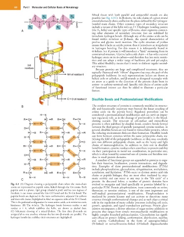
Fig. 6.1 (A) Diagram showing a polypeptide chain where the main-chain evolved to recognize and bind specifically to proteins labeled by a
atoms are represented as peptide units, linked through the Cα atoms. Each particular PTM. Protein phosphorylation, most commonly on serine,
peptide unit is a planar, rigid group (shaded in pink) and has two degrees of threonine, or tyrosine residues, is one of the most important and
freedom; it can rotate around the Cα–CO bond and the N–Cα bond. The well-studied posttranslational modifications. Phosphorylation is
peptide bonds are depicted in the trans conformation: adjacent Cα carbons mediated by protein kinases and can activate or deactivate many
and their side chains (highlighted in blue) on opposite sides of the N–C bond. enzymes through conformational changes and as such plays a critical
This is the preferred configuration for most amino acids, as it minimizes steric role in the regulation of many cellular processes including cell cycle,
hindrance. (B) The α-helix. The hydrogen bonds between residue n and growth, apoptosis, and signal transduction pathways. Protein glyco-
residue n + 4, which stabilizes the helix, are shown as dashed lines. sylation encompasses a diverse selection of sugar-moiety additions to
(C) Schematic drawing of a mixed β-sheet. The first three β-strands are proteins that ranges from simple monosaccharide modifications to
antiparallel to one another, whereas the last two β-strands are parallel. The highly complex branched polysaccharides. Glycosylation has signifi-
hydrogen bonds that stabilize their structures are highlighted. cant effects on protein folding, conformation, distribution, stability,
and activity. Carbohydrates in the form of asparagine-linked
(N-linked) or serine/threonine-linked (O-linked) oligosaccharides