
Page 976 - Hematology_ Basic Principles and Practice ( PDFDrive )
P. 976
Chapter 57 Pharmacology and Molecular Mechanisms of Antineoplastic Agents for Hematologic Malignancies 859
monomer and is concentrated in nucleoli, although smaller amounts recognition and binding (curved and supercoiled DNA, as well as
are found in a diffuse nuclear distribution. The gene for this enzyme DNA crossovers, are preferred), the sequential cleavage of the two
is located on human chromosome 20q12–13.2. Topoisomerase I does strands of DNA with covalent attachment of a monomer to each
not require ATP for catalytic activity. It binds double-strand DNA 5′-end of the cleaved DNA, passage of another DNA duplex through
over 15–25 bp (with a preference for supercoiled or bent DNA) the break site (e.g., to relieve DNA torsional strain or decatenate
followed by cleavage of one DNA strand and forming a transient daughter chromosomes at the end of replication), religation of the
covalent phosphotyrosyl bond at the 3′-end of DNA. DNA torsional cleaved DNA, and ATP hydrolysis-dependent enzyme turnover.
strain is then relieved by a “controlled rotation” mechanism (see Fig. The binding of ATP by topoisomerase II is required for the strand
57.1), subsequent to which the cleaved DNA is religated. The three- passage reaction. Again, the association between topoisomerase II
dimensional crystal structure of human topoisomerase I, both in monomers and the 5′-end of the cleaved DNA has been termed the
covalent and noncovalent complexes with DNA, has defined the cleavable complex, the stabilization of which generally correlates with
structural elements of the enzyme that contacts DNA. The associa- the cytotoxic activity of specific topoisomerase II inhibitors.
tion between topoisomerase I and the 3′-end of cleaved DNA has Because topoisomerase I and II inhibitors convert their respective
been termed the cleavable complex, which is stabilized by topoisom- enzymes into DNA-damaging agents, it is usually true that the more
erase I inhibitors. enzyme target a cell contains (provided it is in the nucleus), the more
cytotoxic is the specific inhibitor. An exception to this generalization
is CLL cells, which have abundant topoisomerase I but are not very
DNA Topoisomerase II sensitive to topoisomerase I inhibitors because topoisomerase I
inhibitors are S-phase specific and CLL cells have very few cells in S
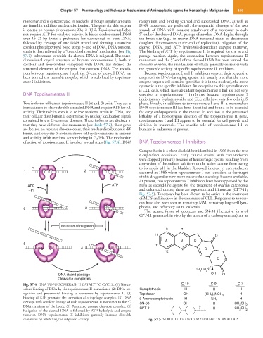
Two isoforms of human topoisomerase II (α and β) exist. They act as phase. Finally, in addition to topoisomerase I and II, a mammalian
homodimers to cleave double-stranded DNA and require ATP for full DNA topoisomerase III has been described and found to be essential
activity. Their role in vivo is to relieve torsional strain in DNA, and for early embryogenesis in the mouse. In addition to the presumed
their cellular distribution is determined by nuclear localization signals lethality of a homozygous deletion of the topoisomerase II gene,
contained in the C-terminal domain. These isoforms are distinct in topoisomerases I and III appear to be essential for cell growth and
that they have different-size monomers (see Table 57.2), their genes division in mammals. The specific role of topoisomerase III in
are located on separate chromosomes, their nuclear distribution is dif- humans is unknown at present.
ferent, and only the α-isoform shows cell cycle variations in amount
and activity (with maximal activity being in G 2 /M). The mechanism
of action of topoisomerase II involves several steps (Fig. 57.4): DNA DNA Topoisomerase I Inhibitors
Camptothecin is a plant alkaloid first identified in 1966 from the tree
Camptotheca acuminata. Early clinical studies with camptothecin
were stopped primarily because of hemorrhagic cystitis resulting from
conversion of the sodium salt form to the active lactone form owing
to its acidic pH in the bladder. Renewed interest in camptothecin
1 2 occurred in 1985 when topoisomerase I was identified as the target
of this drug and as new more water-soluble analogs became available.
At present, two topoisomerase I inhibitors have been approved by the
ADP ATP FDA as second-line agents for the treatment of ovarian carcinoma
+Pi and colorectal cancer; these are topotecan and irinotecan (CPT-11;
Fig. 57.5). Topotecan has been shown to be active in the treatment
of MDS and inactive in the treatment of CLL. Responses to topote-
can have also been seen in refractory MM, refractory large-cell lym-
6 3 phoma, and refractory acute leukemia.
The lactone forms of topotecan and SN-38 (the active form of
CPT-11 generated in vivo by the action of a carboxylesterase) are as
Inhibition of religation
9 7
10 O
A B C N
5 11
4 N D
12
E O
CH CH
3 2
DNA strand passage OH O
Cleavable complexes
Fig. 57.4 DNA TOPOISOMERASE II CATALYTIC CYCLE. (1) Nonco- C-10 C-9 C-7
valent binding of DNA by the topoisomerase II homodimer. (2) DNA rec- Camptothecin H H H
ognition and preferential binding to crossovers by topoisomerase II. (3) Topotecan OH (CH ) NCH 2 H
3 2
Binding of ATP promotes the formation of a topologic complex. (4) DNA 9-Aminocamptothecin H NH 2 H
cleavage with covalent linkage of each topoisomerase II monomer to the 5′- SN-38 OH H CH CH
3
DNA terminus of the break. (5) Poststrand passage cleavable complex. (6) CPT-11 O H CH CH 2
Religation of the cleaved DNA is followed by ATP hydrolysis and enzyme N N C O 3 2
turnover. DNA topoisomerase II inhibitors generally increase cleavable
complexes by inhibiting the religation activity. Fig. 57.5 STRUCTURE OF CAMPTOTHECIN ANALOGS.