
Page 975 - Hematology_ Basic Principles and Practice ( PDFDrive )
P. 975
858 Part VII Hematologic Malignancies
cell transplantation. It is recognized for its immunosuppressive func- totic proteins such as Bcl-X(L), Mcl-1, and Bax with dephosphoryla-
tion and inducing tolerance to allografts transplantation. Fludarabine tion of akt; and inhibits DNA synthesis.
phosphate is the 2-fluoro, 5′-monophosphate derivative of vidarabine It appears more active against B-cell than T-cell lymphomas, but
(9′-β-D-arabinofuranosyladenine [ara-A]) and is converted to the also has activity in AML and myelodysplastic syndromes (MDS). It
di- and triphosphate by intracellular kinases, as are gemcitabine and is well tolerated. Recent studies show its significant efficacy in ara-C–
ara-C. It has greater potency because it confers resistance to deamina- refractory AML. Reversible liver toxicity and myelosuppression can
tion by adenosine deaminase (ADA) and improved solubility. It is be dose limiting (see Chapter 59).
incorporated into DNA as a nucleotide and causes chain termination.
It is a potent inhibitor of cytosolic 5′-nucleotidase II. It is also a Nelarabine
substrate for uracil glycosylase, causing abasic sites as the first stem Nelarabine (9-β-D-arabinofuranosylguanine) is another FDA-
in BER and has recently been used in combination with TRC102, approved purine analog for the treatment of refractory T-cell leuke-
which binds to these sites and prevents their repair. Like other mias and lymphomas (Chapter 85).
nucleoside analogues, it causes replication fork collapse, double- As a nucleotide analogue, it preferentially accumulates in T cells
strand breaks, and induction of P53, leading to apoptotic signaling. and is incorporated into DNA, causing chain termination and
Its efficacy against normal lymphoid T and B cells appears linked to inhibiting DNA synthesis. The FDA approved this drug after analyz-
both cytotoxicity against proliferating cells and resting cells; the latter ing the results of two phase II clinical trials, one in pediatric T-cell
effect is mediated by interference with the normal activity of the acute lymphoblastic leukemia (ALL) and the other in adults with
nucleotide excision repair pathway. T-cell lymphoblastic lymphoma. In both cases, patients had relapsed
Fludarabine is a complex agent. Resistance is multifactorial after at least two induction regimens. Because CRs were seen in 13%
including active transport, decreased cytosolic 5′-nucleotidase II and of the 39% of pediatric patients and in 18% of the 28 adult patients,
deoxycytokine kinase, and Ku80 binding to telomerase. Other non- the FDA granted approval. Neurologic toxicity is dose limiting. Good
specific mechanisms include mutations in p53 or loss due to chromo- response rates, including CRs, have been seen in patients with refrac-
some deletion, which is important in many cases of CLL, and other tory T-cell leukemias.
proliferation-associated genes such as NOTCH1, SF3B1, and BIRC3. Reduced ara-G incorporation into DNA is likely due to both
Low-level miR-34a is also associated with fludarabine resistance. altered nucleotide transport, increased nucleotidase activity, reduced
2
Fludarabine, at a standard dose of 25 mg/m for 5 days and nucleoside kinase activity, and nonspecific resistance mechanisms as
rituximab with or without cyclophosphamide were the mainstay of described earlier for this class of agent.
primary treatment for CLL (Chapter 77). Fludarabine has also been
used in refractory leukemias, marginal cell and other low-grade
lymphomas. Recognition of the appearance of lymphopenia after Inhibitors of DNA Topoisomerase I and II
treatment led to studies establishing fludarabine as part of the non-
myelosuppressive preparative regimens for allogeneic transplantation, Inhibitors of DNA topoisomerases I and II include such drugs as
used particularly for older individual recipients. This is most often at doxorubicin, daunorubicin, mitoxantrone, etoposide, and topotecan.
2
a dose of 30–35 mg/m for 5 days and used in combination with Before describing the specific inhibitors, a brief review of the drug
radiation, melphalan, or cyclophosphamide. In addition to lympho- targets (topoisomerase enzymes) will be presented (Table 57.2).
penia, multiple cycles are associated with prolonged myelosuppression
and a chronic peripheral neuropathy.
DNA Topoisomerase I
Clofarabine
Clofarabine (2-chloro-2′-arabino-flouro-2′-deoxyadenosine) is a Topoisomerase I is a ubiquitous enzyme whose function in vivo is to
purine analog with activity in patients with relapsed acute leukemia. relieve the torsional strain in DNA, specifically to remove positive
Its activation requires cellular uptake and conversion to the triphos- supercoils generated in front of the replication fork and to relieve
phate nucleotide. It then decreases ribonucleotide reductase; alters negative supercoils occurring downstream of RNA polymerase during
nucleotide precursors; inhibits and reduces the function of antiapop- transcription. Topoisomerase I is catalytically active as a 100-kDa
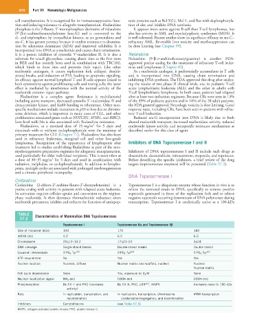
TABLE Characteristics of Mammalian DNA Topoisomerases
57.2
Topoisomerase I Topoisomerase IIα and Topoisomerase IIβ
Size of monomer (kDa) 100 170 180
mRNA (kb) 4.2 6.2 6.5
Chromosome 20q12–13.2 17q21–22 3p24
DNA cleavage Single-strand breaks Double-strand breaks Double strand
Covalent intermediate 3′PO 4 -Tyr 723 5′PO 4 -Tyr 804 5′PO 4 -Tyr 821
ATP requirement No Yes Yes
Nuclear location Nucleoli, diffuse Nuclear matrix and scaffold, nucleoli Nucleoli
Nuclear matrix
Cell cycle dependence None Yes, maximum in G 2 /M None
Nuclear localization signal NH 2 -end COOH-end COOH-end
Phosphorylation By CK II and PKC (increases By CK II, PKC, p34 odc2 , MAPK Increases mass to 190 kDa
activity)
Role In replication, transcription, and In replication, transcription, chromosome rRNA transcription
recombination condensation/segregation, and recombination
Inhibitors Camptothecins (see Table 57.3)
MAPK, mitogen-activated protein kinase; PKC, protein kinase C.