
Page 1040 - Williams Hematology ( PDFDrive )
P. 1040
1014 Part VII: Neutrophils, Eosinophils, Basophils, and Mast Cells Chapter 66: Disorders of Neutrophil Function 1015
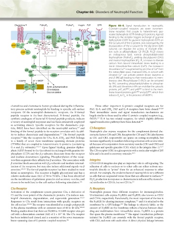
Figure 66–4. Signal transduction in neutrophils.
G-protein–coupled receptors are seven transmem-
brane receptors that couple to heterotrimeric gua-
nosine triphosphate (GTP)-binding (G) proteins. Agonist
binding to the receptor triggers exchange of guanine
diphosphate (GDP) for guanine triphosphate (GTP) on
the Gα subunit of the G-protein, and consequently, the
disassociation of the α subunit for the βg-dimer. Both
subunits can regulate the activity of multiple effec-
tors such as phospholipase Cβ (PLCβ). PLCβ cleaves
an endogenous lipid, namely phosphatidylinositol
bisphosphate (PtdInsP ), yielding diacylglycerol (DAG)
2
and inositol trisphosphate (IP ). IP is known to liberate
3
3
calcium from bound intracellular stores leading to a
rise in intracellular-free calcium (Ca )i. The increase in
2+
2+
intracellular Ca is augmented by an influx of Ca from
2+
the extracellular space. Increased DAG, in concert with
elevated Ca can activate protein kinase isozymes α
2+
and β (PKCαβ) leading to their translocation to mem-
branous sites. Phospholipase D (PLD) can be activated
by PKC converting phosphatidylcholine to phosphat-
idic (PA) acid. Elevations in PA can mobilize the cytosolic
proteins, p47, p67 phox , and p40 phox to bind to the mem-
brane-bound proteins gp91 phox and p22 phox , which then
reduces O to O in the presence of NADPH.
2 2–
chemokine and chemotactic factors produced during the inflamma- Three other important G-protein–coupled receptors are for
tory process activate neutrophils by binding to specific cell-surface PAF, IL-8, and LTB . PAF and IL-8 receptors have been cloned. 227,228
4
receptors. Of the neutrophil chemotactic receptors, the N-formyl Their intracellular stores and signal transduction mechanisms are
peptide receptor is the best characterized. N-formyl peptide, the largely similar to those used by other G-protein–coupled receptors (e.g.,
synthetic analogues of bacterial N-formyl peptide products, induces fMLP). IL-8 has two related receptors, for which slightly different
227
a variety of neutrophil responses and has been extensively employed signal transduction pathways have been detected. 229
as activating stimuli. Specific receptors for the chemotactic pep-
tide, fMLP, have been identified on the neutrophil surface, and C3 Receptors
216
binding of the formyl peptide to its receptor correlates with its abil- Neutrophils also express receptors for the complement-derived che-
ity to induce chemotaxis and degranulation. The formyl peptide motactic factors C3b and C3bi. Receptors for C3b and C3bi (also known
217
receptor, like the receptors for C5a, IL-8, LTB , and PAF, belongs as CR1 and CR3, respectively) are sparse on resting neutrophils, but
218
4
to a family of seven trans-membrane–spanning domain proteins increase significantly in numbers following activation with several stim-
(7TMRs) that are coupled to heterotrimeric G-proteins (containing uli because of incorporation from secretory vesicles (CR1 and CR3) and
G α and β,γ subunits). 219,213 Upon ligand binding, guanine diphos- gelatinase and specific granules (CR3, which is the integrin Mac-1). 32,71
phate (GDP) bound to the Gα subunit is exchanged with guanine tri- The C3b receptor (CR1) is a glycoprotein with a molecular weight of 205
phosphate (GTP) and the β,γ subunits dissociate from the receptor kDa and is located in secretory vesicules. 65,71
and mediate downstream signaling. Phosphorylation of the recep-
tors then augments their affinity for β arrestins. The association with Integrins
β arrestins block association with β,γ subunits and mediate internal- CD11/CD18 integrins also play an important role in cell signaling. The
ization of the receptors, but may also induce additional signals via β adhesion of cells to surfaces or to other cells can either activate neu-
arrestins. 220,221 The formyl peptide receptor has been studied in most trophils directly or “prime” them for an enhanced response to other
detail in neutrophils. The receptor is highly glycosylated and has a stimuli. For example, the oxidative burst of neutrophils is very different
relative molecular mass (Mr) of 50 to 70 kDa. It has been identified in cells that are suspended versus those that are adherent to surfaces.
230
on the membranes of gelatinase granules and secretory vesicles, and H O production in response to chemotaxins is influenced by monoclo-
2
2
shown to be mobilized to the cell surface following stimulation. 222 nal antibodies to CD11b, but not CD11a. 231
C5a Receptor Fc Receptors
Activation of the complement system generates C5a, a derivative of Neutrophils possess three different receptors for immunoglobulins.
C5 and the most potent of the chemotactic proteins. C5a induces neu- Unstimulated cells express FcγRIIA and FcγRIII, also known as CD32
trophil chemotaxis, degranulation, and superoxide generation. 222,223 and CD16, respectively. Functionally, the most important of the two is
Responses to C5a result from interactions with specific receptors on the FcγRIII for clearing immune complexes, and it is attached to the
232
the cell surface. 222,224 The receptor was identified as a single polypeptide membrane by a GPI linkage. The linkage is relatively labile, so the
232
in the plasma membrane with an apparent mass of 40 to 48 kDa. 222,225 amount of FcγRIII on the membrane reflects a balance between shed-
Binding studies show that there are 50,000 to 113,000 receptor sites per ding and mobilization from intracellular stores. FcγRIIA is a protein
cell with a dissociation constant (Kd) of 2 × 10 M. The C5a receptor that spans the plasma membrane. The signal transduction pathways
–9
233
has been isolated and cloned, and is a member of the seven transmem- initiated by FcγRIII can crosstalk with the formyl peptide receptor,
brane-spanning class of G-protein–coupled receptors. 226 with CR3, and even with each other. A direct physical linkage between
Kaushansky_chapter 66_p1005-1042.indd 1015 9/21/15 10:48 AM